
In the study of anatomy, one of the most fascinating and complex organs to dissect is the heart. A sheep heart, in particular, is commonly used for dissections due to its similarities to a human heart. This worksheet provides answers to questions that students may encounter during their sheep heart dissection.
The sheep heart dissection worksheet answers provide a step-by-step guide on how to identify and dissect the different parts of the heart, such as the coronary arteries, ventricles, and atria. It also explains the functions of each part and their role in the circulatory system.
By following the worksheet answers, students can gain a deeper understanding of the anatomy and physiology of the heart. They can learn about the importance of the heart in pumping blood throughout the body, the structure of the heart valves, and the significance of the electrical impulses that regulate the heartbeat.
Overall, the sheep heart dissection worksheet answers serve as an educational resource to enhance students’ knowledge and understanding of the heart. Through hands-on dissection and the guidance provided in the worksheet, students can explore the intricacies of this vital organ and appreciate its role in maintaining the body’s overall health and well-being.
Sheep Heart Dissection Worksheet Answers
In a sheep heart dissection worksheet, students are typically asked to identify and label the different structures and chambers of the heart. It is a hands-on activity that allows students to explore the anatomy of the heart and gain a better understanding of how it functions.
Here are some possible answers to a sheep heart dissection worksheet:
- Atria: The two upper chambers of the heart, the left and right atria, receive blood from the veins and pump it into the ventricles.
- Ventricles: The two lower chambers of the heart, the left and right ventricles, receive blood from the atria and pump it out to the rest of the body.
- Aorta: The largest artery in the body, the aorta carries oxygenated blood from the left ventricle to the rest of the body.
- Pulmonary artery: The artery that carries deoxygenated blood from the right ventricle to the lungs for oxygenation.
- Pulmonary veins: The veins that carry oxygenated blood from the lungs to the left atrium.
- Tricuspid valve: Located between the right atrium and right ventricle, the tricuspid valve prevents the backflow of blood into the atrium when the ventricle contracts.
- Bicuspid valve: Also known as the mitral valve, the bicuspid valve is located between the left atrium and left ventricle and prevents the backflow of blood.
- Semilunar valves: These valves, including the pulmonary and aortic valves, prevent the backflow of blood from the arteries to the ventricles.
By completing a sheep heart dissection and answering the worksheet questions, students can gain a deeper understanding of the internal structures and functions of the heart. This activity is an important part of studying the circulatory system and can help students grasp the complexity of the human cardiovascular system.
Materials and Tools Required for the Dissection
When performing a sheep heart dissection, it is essential to have the necessary materials and tools ready. These items will help ensure a successful and efficient dissection process. Here is a list of some of the materials and tools that are commonly used:
Materials:
- Sheep heart (fresh or preserved)
- Dissection tray or cutting board
- Scalpel or dissection scissors
- Forceps
- Dissection pins
- Dissection gloves
- Plastic or glass container for fluid waste
- Dissection worksheet or guide
Tools:
- Dissection microscope (optional, for a more detailed analysis)
- Scalpel blades (if using a scalpel)
- Dissection probe or blunt probe
These materials and tools are essential for a successful dissection. The sheep heart is the main specimen that will be dissected, and it can be obtained from a local butcher or a supplier specializing in biological specimens. The dissection tray or cutting board provides a stable and clean surface to work on, preventing contamination. The scalpel or dissection scissors are used to make precise incisions and cut through tissue. Forceps are helpful in holding and manipulating the heart during the dissection process.
Dissection pins are commonly used to hold back flaps of tissue, providing a clear view of the internal structures. Dissection gloves are important for personal protection and maintaining a sterile environment. A plastic or glass container is necessary for disposing of fluid waste generated during the dissection. A dissection worksheet or guide provides step-by-step instructions and can be used to record observations and findings during the dissection.
In some cases, a dissection microscope may be used for a more detailed analysis of the heart structures. Scalpel blades may be required if using a scalpel that needs blade replacements. Finally, a dissection probe or blunt probe can be helpful in separating tissues or exploring specific structures.
External Anatomy of the Sheep Heart
The external anatomy of the sheep heart reveals several distinct features that play crucial roles in its function as a vital organ. The sheep heart is located in the thoracic cavity, just behind the sternum and between the lungs. It is generally conical in shape, with a pointed apex at the bottom and a broad base at the top. The heart is surrounded by a double-layered protective sac called the pericardium.
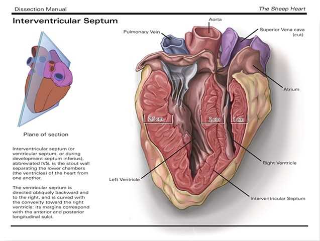
One of the most noticeable features of the sheep heart is the four chambers that make up its structure. The lower two chambers, known as the ventricles, are responsible for pumping blood out to the body and lungs. The upper two chambers, called the atria, receive blood from different parts of the body and lungs. The division between the atria and ventricles is marked by a thick wall known as the septum, which ensures that oxygen-rich and oxygen-poor blood do not mix.
- Atria: The left atrium receives oxygenated blood from the lungs, while the right atrium receives deoxygenated blood from the body.
- Ventricles: The left ventricle pumps oxygenated blood to the body, while the right ventricle pumps deoxygenated blood to the lungs.
- Aortic Arch: This curved vessel arises from the left ventricle and carries oxygenated blood to the rest of the body.
- Pulmonary Artery: This artery arises from the right ventricle and carries deoxygenated blood to the lungs for oxygenation.
- Pulmonary Veins: These veins carry oxygenated blood from the lungs to the left atrium.
- Vena Cavae: There are two vena cavae – the superior vena cava, which carries deoxygenated blood from the upper body to the right atrium, and the inferior vena cava, which carries deoxygenated blood from the lower body to the right atrium.
The external anatomy of the sheep heart provides a clear understanding of its structure and function. Each component, from the chambers to the blood vessels, has a specific role in ensuring the continuous flow of oxygenated and deoxygenated blood throughout the body. Through dissection and exploration, we can gain valuable insights into the intricate workings of this remarkable organ.
Internal Anatomy of the Sheep Heart
The sheep heart is a complex organ that plays a vital role in the circulatory system. It is composed of various structures that work together to pump and distribute blood throughout the body. Understanding the internal anatomy of the sheep heart is essential for studying its function and understanding its role in the cardiovascular system.
1. Chambers: The sheep heart is divided into four chambers – two atria and two ventricles. The atria are the upper chambers, while the ventricles are the lower chambers. The right atrium receives deoxygenated blood from the body, while the left atrium receives oxygenated blood from the lungs. The right ventricle pumps deoxygenated blood to the lungs, and the left ventricle pumps oxygenated blood to the rest of the body.
2. Valves: The heart contains several valves that regulate the flow of blood through the chambers. The atrioventricular (AV) valves separate the atria from the ventricles and prevent backflow of blood. The tricuspid valve is located between the right atrium and right ventricle, and the mitral valve is located between the left atrium and left ventricle. The semilunar valves are located at the exits of the ventricles and prevent backflow of blood into the chambers. The pulmonary valve is found in the right ventricle, and the aortic valve is found in the left ventricle.
- The tricuspid valve
- The mitral valve
- The pulmonary valve
- The aortic valve
3. Blood Vessels: The sheep heart is connected to various blood vessels that transport blood to and from the heart. The superior and inferior vena cava bring deoxygenated blood from the upper and lower parts of the body, respectively, to the right atrium. The pulmonary veins carry oxygenated blood from the lungs to the left atrium. The pulmonary artery carries deoxygenated blood from the right ventricle to the lungs for oxygenation. The aorta is the largest artery in the body and carries oxygenated blood from the left ventricle to the rest of the body.
Overall, the internal anatomy of the sheep heart showcases the intricate structures and mechanisms involved in its function as a powerful organ responsible for circulating blood throughout the body. Understanding this anatomy is crucial for studying the cardiovascular system and its role in maintaining overall health and well-being.
Dissection Procedure Step-by-Step
The dissection of a sheep heart is a common laboratory activity that allows students to explore the anatomy and functions of the organ. It is important to follow a step-by-step procedure to ensure that the dissection is conducted safely and effectively. The following steps outline the procedure for dissecting a sheep heart:
Step 1: Preparation
Before beginning the dissection, gather all the necessary materials, including a preserved sheep heart, dissecting tray, dissecting tools (scalpel, scissors, and forceps), gloves, and a lab coat or apron. Ensure that the work area is clean and well-ventilated.
Step 2: External Examination
Start by placing the sheep heart on the dissecting tray and carefully examine its external features. Observe the size, shape, and color of the heart. Identify the major blood vessels attached to the heart, including the aorta, pulmonary artery, and vena cava.
Step 3: Opening the Heart

Using a scalpel, make a midline incision through the pericardium surrounding the heart. Be cautious not to cut too deep to avoid damaging the underlying structures. Gently remove the pericardium, exposing the surface of the heart.
Step 4: Internal Examination
Take a closer look at the internal structures of the heart. Locate the four chambers: the right atrium, right ventricle, left atrium, and left ventricle. Observe the valves, such as the tricuspid valve and the mitral valve, which separate the chambers. Examine the walls of the heart and identify the coronary arteries.
Step 5: Dissecting the Blood Vessels
To examine the blood vessels, carefully cut away the major vessels connected to the heart. Identify the aorta and trace it to its branches. Observe the pulmonary artery and the veins entering the heart. Take note of any abnormalities or variations in the vessel structures.
Step 6: Clean up
Once the dissection is complete, clean up the dissecting tray and tools. Dispose of any waste materials in the appropriate manner. Wash your hands thoroughly to remove any residue or chemicals.
By following this step-by-step procedure, students can gain a better understanding of the intricacies of a sheep heart and its vital role in circulation and oxygenation of the body.
Observations and Findings during the Dissection
During the sheep heart dissection, several observations and findings were made that helped us understand the structure and function of the heart.
External features: The sheep heart was approximately the size of a human fist and had a conical shape. It was covered by a protective layer called the pericardium. The heart was divided into four chambers: two atria and two ventricles. The heart also had several blood vessels attached to it, such as the superior and inferior vena cava, pulmonary artery, and aorta.
Internal features: Upon opening the heart, we observed the partitions that separated the atria and ventricles called the interatrial and interventricular septa, respectively. The heart valves, including the tricuspid valve, bicuspid valve (also known as the mitral valve), pulmonary valve, and aortic valve, were clearly visible. These valves ensured the one-way flow of blood through the heart.
Blood flow: As we followed the blood flow through the heart, we observed that deoxygenated blood entered the right atrium through the superior and inferior vena cava. From there, it passed through the tricuspid valve into the right ventricle. The right ventricle then pumped the deoxygenated blood through the pulmonary valve and into the pulmonary artery, which leads to the lungs for oxygenation. Oxygenated blood returned to the left atrium through the pulmonary veins. From the left atrium, it passed through the bicuspid valve into the left ventricle. Finally, the left ventricle pumped the oxygenated blood through the aortic valve and into the aorta to be circulated throughout the body.
Structural adaptations: We noticed that the walls of the left ventricle were thicker and more muscular than those of the right ventricle. This is because the left ventricle needs to generate greater force to pump blood throughout the body, whereas the right ventricle only needs to pump blood to the lungs. Additionally, the heart muscle itself had a unique texture, consisting of striated fibers.
Overall function: Through the dissection, we gained a better understanding of how the heart functions as a pump to circulate blood throughout the body. The coordinated contraction and relaxation of the atria and ventricles, along with the opening and closing of the valves, ensure efficient blood flow and oxygenation.