
The anatomy of a synapse worksheet answer key provides a comprehensive understanding of the structure and function of a synapse. A synapse is a specialized junction between two neurons that allows for the transmission of signals in the nervous system. It is a crucial component in the process of information processing and transmission in the brain and plays a vital role in cognition, perception, and behavior.
The worksheet answer key breaks down the anatomy of a synapse into its main components, namely the presynaptic terminal, the synaptic cleft, and the postsynaptic membrane. Each component is examined in detail, highlighting its unique features and functions. The key also provides a clear explanation of how these components work together to facilitate signal transmission.
In addition to the structural aspects, the answer key delves into the molecular and cellular mechanisms underlying synaptic transmission. It elucidates the role of neurotransmitters, such as dopamine, serotonin, and glutamate, in the synaptic cleft and their interaction with specific receptors on the postsynaptic membrane. Furthermore, it discusses the process of synaptic plasticity, which refers to the ability of synapses to change and strengthen their connections over time.
Overall, the anatomy of a synapse worksheet answer key serves as a valuable resource for students and researchers alike. It provides a comprehensive overview of the complex and intricate nature of synapses, shedding light on the fundamental processes that underlie brain function and how disruptions in synaptic communication can give rise to neurological disorders. Understanding the anatomy of a synapse is crucial for unraveling the mysteries of the brain and advancing our knowledge of cognition and behavior.
The Anatomy of a Synapse Worksheet Answer Key

The anatomy of a synapse is a complex structure that plays a crucial role in the communication between neurons in the nervous system. A synapse is the junction between two neurons, where they can transfer information through chemical or electrical signals. This worksheet provides an answer key to help students understand the different components of a synapse and their functions.
1. Synaptic Vesicles – These small sacs contain neurotransmitters, which are chemical messengers that transmit signals between neurons. When an action potential reaches the synaptic terminal, these vesicles fuse with the presynaptic membrane and release their neurotransmitters into the synaptic cleft.
2. Presynaptic Membrane – This is the cell membrane of the presynaptic neuron, which contains voltage-gated calcium channels. These channels open in response to an action potential, allowing calcium ions to enter the synaptic terminal and trigger the release of neurotransmitters.
3. Synaptic Cleft – This is the small gap between the presynaptic membrane and the postsynaptic membrane. After neurotransmitters are released into the synaptic cleft, they diffuse across this gap and bind to receptors on the postsynaptic membrane.
4. Postsynaptic Membrane – This is the cell membrane of the postsynaptic neuron, which contains receptors that can bind to neurotransmitters. When neurotransmitters bind to these receptors, they can either excite or inhibit the postsynaptic neuron, depending on the type of neurotransmitter and the receptor.
5. Reuptake Transporters – These proteins are located on the presynaptic membrane and are responsible for reabsorbing excess neurotransmitters from the synaptic cleft back into the presynaptic neuron. This process helps to terminate the signal and prevent the accumulation of neurotransmitters in the synapse.
- 6. Excitatory and Inhibitory Neurotransmitters – Neurotransmitters can be classified as either excitatory or inhibitory, depending on their effect on the postsynaptic neuron. Excitatory neurotransmitters, such as glutamate, increase the likelihood of the postsynaptic neuron firing an action potential. Inhibitory neurotransmitters, such as GABA, decrease the likelihood of an action potential.
- 7. Neurotransmitter Receptors – These proteins are located on the postsynaptic membrane and bind to specific neurotransmitters. Each receptor is selective for a particular neurotransmitter, allowing for precise communication between neurons. Activation of these receptors can initiate a series of intracellular events, leading to changes in the postsynaptic neuron and ultimately influencing its firing rate.
| Component | Function |
|---|---|
| Synaptic Vesicles | Contain neurotransmitters for release |
| Presynaptic Membrane | Contains calcium channels for neurotransmitter release |
| Synaptic Cleft | Gap between pre- and postsynaptic membranes |
| Postsynaptic Membrane | Contains neurotransmitter receptors |
| Reuptake Transporters | Reabsorb excess neurotransmitters |
In conclusion, the anatomy of a synapse is a complex and organized structure that allows for the transmission of signals between neurons. Understanding the different components of a synapse and their functions is essential in grasping how information is processed and communicated in the nervous system.
Overview of Synapses
A synapse is a specialized connection between two neurons that allows for communication and the transfer of information. It is the fundamental unit of communication in the nervous system, enabling the transmission of signals between neurons and ultimately controlling various functions of the body.
Synapses are essential for the proper functioning of the nervous system. They are responsible for the formation and modification of neural circuits, which are the basis of learning and memory. Without synapses, the transmission of information between neurons would not be possible, and the complexity of the brain’s processing abilities would be severely limited.
Structure of a Synapse
A synapse consists of three main components: the presynaptic neuron, the postsynaptic neuron, and the synaptic cleft. The presynaptic neuron is the sending neuron, which releases neurotransmitters into the synaptic cleft. The postsynaptic neuron is the receiving neuron, which detects the neurotransmitters and initiates an electrical impulse. The synaptic cleft is the small gap between the two neurons where the neurotransmitters diffuse.
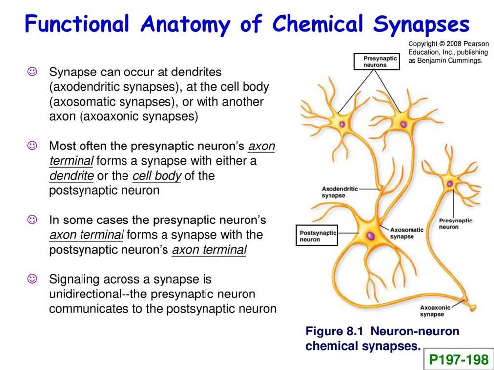
Types of Synapses
Synapses can be categorized into two main types: electrical synapses and chemical synapses. Electrical synapses are characterized by the direct flow of ions between neurons through gap junctions, allowing for rapid and bidirectional communication. On the other hand, chemical synapses involve the release of neurotransmitters into the synaptic cleft, which bind to receptors on the postsynaptic neuron to initiate an action potential.
Function of Synapses
The primary function of synapses is to transmit and integrate electrical signals between neurons. When an action potential reaches the presynaptic neuron, it triggers the release of neurotransmitters, which bind to receptors on the postsynaptic neuron, causing depolarization and the generation of a new action potential. This process allows for the propagation of electrical signals throughout the nervous system, enabling the coordination of various physiological and behavioral responses.
| Key Points |
|---|
| Synapses are specialized connections between neurons that enable communication and the transfer of information. |
| They consist of the presynaptic neuron, postsynaptic neuron, and synaptic cleft. |
| Synapses can be electrical or chemical, depending on the type of communication they facilitate. |
| The main function of synapses is to transmit and integrate electrical signals between neurons, allowing for the coordination of various physiological and behavioral responses. |
Structure of a Synapse
A synapse is a structure that allows neurons to communicate with each other. It consists of three main components: the presynaptic terminal, the synaptic cleft, and the postsynaptic membrane. These components work together to transmit signals between neurons in the brain and nervous system.
The presynaptic terminal is the end of the neuron that releases neurotransmitters. It contains vesicles, which store the neurotransmitters, and mitochondria, which provide energy for the release of these chemical messengers. The presynaptic terminal also has voltage-gated calcium channels, which are essential for triggering the release of neurotransmitters into the synaptic cleft.
The synaptic cleft is a narrow gap between the presynaptic terminal and the postsynaptic membrane. It is filled with extracellular fluid that helps facilitate the diffusion of neurotransmitters. The distance of this gap is only about 20 to 50 nanometers, allowing for efficient communication between neurons.
The postsynaptic membrane, on the other hand, is the receiving end of the synapse. It contains specialized proteins called receptors that bind to the neurotransmitters released by the presynaptic terminal. These receptors can be ion channels, which allow ions to flow into or out of the cell, or they can activate signaling pathways within the postsynaptic neuron.
In summary, the structure of a synapse is crucial for the transmission of signals between neurons. The presynaptic terminal releases neurotransmitters into the synaptic cleft, which then bind to receptors on the postsynaptic membrane, resulting in the generation of an electrical signal in the postsynaptic neuron. Understanding the anatomy of a synapse is essential for understanding how neurons communicate and how the brain functions as a whole.
Presynaptic Neuron
The presynaptic neuron is a crucial component of the synapse, which is the point of communication between two neurons. It is responsible for transmitting signals to the postsynaptic neuron, allowing for the transmission of information throughout the nervous system. The presynaptic neuron plays a vital role in the overall functioning of the brain and is involved in various physiological processes.
At the synapse, the presynaptic neuron releases neurotransmitters, which are chemical messengers that transmit signals from one neuron to another. These neurotransmitters are stored in vesicles within the presynaptic terminal. When an action potential reaches the presynaptic terminal, it triggers the opening of calcium channels, allowing calcium ions to enter the terminal. This influx of calcium ions leads to the release of neurotransmitters into the synaptic cleft.
The presynaptic neuron is highly specialized in its function, with unique structures and mechanisms that facilitate neurotransmitter release. For example, it contains a variety of proteins, such as synaptic vesicle proteins and transporters, that are involved in the packaging and release of neurotransmitters. Additionally, the presynaptic terminal is enriched in mitochondria, which provide the energy needed for neurotransmitter synthesis and transport.
- The presynaptic neuron plays a crucial role in transmitting signals to the postsynaptic neuron.
- It releases neurotransmitters into the synaptic cleft.
- It contains specialized structures and proteins that facilitate neurotransmitter release.
- The presynaptic terminal is enriched in mitochondria for energy production.
In summary, the presynaptic neuron is a key player in synaptic transmission. Its ability to release neurotransmitters and transmit signals is essential for the proper functioning of the nervous system. Understanding the anatomy and function of the presynaptic neuron is crucial for gaining insight into the complex mechanisms underlying synaptic communication.
Postsynaptic Neuron
The postsynaptic neuron is the target neuron that receives signals from the presynaptic neuron. It plays a crucial role in the transmission of information between neurons in the brain and nervous system. The postsynaptic neuron contains specialized structures called dendrites, which receive neurotransmitter molecules released by the presynaptic neuron.
Dendrites: Dendrites are branch-like structures that extend from the cell body of the postsynaptic neuron. They have numerous projections called dendritic spines, which provide additional surface area for synapses to form. The dendrites receive neurotransmitters from the presynaptic neuron and relay the electrical signals to the cell body.
The postsynaptic neuron also has a cell body, which contains the nucleus and other organelles necessary for cellular functions. From the cell body, the electrical signals are transmitted through the axon, a long fiber-like structure that carries the signals to other neurons or effector cells.
Postsynaptic Density: The postsynaptic density is a region in the postsynaptic neuron where the neurotransmitter receptors and signaling proteins are concentrated. It is responsible for the amplification and integration of the synaptic signals. The postsynaptic density contains various proteins and receptors that bind with specific neurotransmitters, which initiate a cascade of chemical reactions leading to the generation of electrical signals.
Synaptic Plasticity: Synaptic plasticity refers to the ability of the synapse to change its strength and connectivity in response to activity and experience. It plays a vital role in learning and memory formation. The postsynaptic neuron is involved in synaptic plasticity by modulating the strength of the synapse through various mechanisms, such as the insertion or removal of receptors and changes in the number and location of synapses.
In summary, the postsynaptic neuron is a crucial component of the synapse and plays an essential role in receiving and integrating signals from the presynaptic neuron. The dendrites and postsynaptic density are critical structures for signal transmission, and synaptic plasticity allows for the modulation of synaptic strength and connectivity for effective brain function.
Synaptic Transmission
Synaptic transmission is the process by which neurons communicate with each other through chemical signals at specialized junctions called synapses. These synapses are the key functional units of the nervous system, allowing for the transmission of information from one neuron to another.
The process of synaptic transmission begins with the arrival of an action potential, or electrical signal, at the presynaptic terminal of a neuron. This electrical signal triggers the release of neurotransmitters, which are chemical messengers stored in vesicles within the presynaptic terminal. The neurotransmitters are then released into the synaptic cleft, the small gap between the presynaptic and postsynaptic neurons.
At the postsynaptic neuron, the neurotransmitters bind to specific receptors on the surface of the neuron. These receptors are typically ion channels, allowing ions to flow into or out of the postsynaptic neuron. This produces a change in the electrical potential of the postsynaptic membrane, either depolarizing or hyperpolarizing it.
The change in electrical potential, known as a synaptic potential, can be excitatory or inhibitory. If the synaptic potential depolarizes the postsynaptic membrane, bringing it closer to the threshold for generating an action potential, it is considered excitatory. Conversely, if the synaptic potential hyperpolarizes the postsynaptic membrane, making it less likely to generate an action potential, it is considered inhibitory.
The strength of the synaptic transmission can be modulated by various factors, such as the amount of neurotransmitters released, the number and sensitivity of receptors, and the efficiency of neurotransmitter reuptake or degradation. These factors contribute to the fine-tuning of neural communication, allowing for precise control of information processing in the brain.
Summary:
- Synaptic transmission is the process of communication between neurons through chemical signals at synapses.
- An action potential triggers the release of neurotransmitters from the presynaptic neuron into the synaptic cleft.
- Neurotransmitters bind to receptors on the postsynaptic neuron, producing a change in the electrical potential.
- Synaptic potentials can be excitatory or inhibitory, depending on their effect on the postsynaptic membrane potential.
- The strength of synaptic transmission can be modulated by various factors, allowing for precise control of information processing in the brain.