
The EKG LabQuest 12 is a cutting-edge device used to analyze the electrical activity of the heart. This state-of-the-art technology provides healthcare professionals with valuable insights into the heart’s function and can aid in the diagnosis and treatment of various cardiovascular conditions.
With EKG LabQuest 12, medical experts can measure the electrical signals generated by the heart and detect any abnormalities or irregularities in its rhythm and structure. By analyzing these signals, healthcare professionals can assess the overall health of the heart and identify potential problems such as arrhythmias, conduction disorders, and ischemia.
One of the key benefits of using EKG LabQuest 12 is its accuracy and precision. The device captures high-quality data, allowing medical professionals to make informed decisions regarding patient care. Additionally, EKG LabQuest 12 offers advanced analysis tools and features that enable healthcare providers to interpret the collected data more effectively and make accurate diagnoses.
Overall, EKG LabQuest 12 is a crucial tool in modern cardiology, providing healthcare professionals with a comprehensive understanding of the heart’s electrical activity. Its advanced features and accurate measurements make it an invaluable asset in diagnosing and treating various cardiac conditions, ultimately improving patient outcomes and quality of life.
Analyzing the Heart with EKG Lab Quest 12 Answers
The EKG Lab Quest 12 is a valuable tool for analyzing the heart’s electrical activity and diagnosing various heart conditions. By connecting electrodes to the body and recording the electrical signals produced by the heart, healthcare professionals can gain important insights into the heart’s performance. In this lab, students learn to interpret EKG waveforms and identify abnormalities that may indicate cardiac issues.
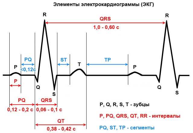
One of the key answers provided by this lab is the ability to identify different components of an EKG waveform. The waveform consists of several distinct segments and intervals, such as the P wave, the PR interval, the QRS complex, and the T wave. By understanding the significance of each of these components, medical professionals can diagnose conditions like atrial fibrillation, ventricular hypertrophy, or myocardial infarction.
Another important aspect of the EKG Lab Quest 12 answers is the recognition of arrhythmias. Arrhythmias refer to abnormal heart rhythms that can be potentially dangerous if left untreated. By analyzing the EKG waveforms and identifying irregularities in the heart’s electrical activity, medical professionals can diagnose and treat various types of arrhythmias, including atrial fibrillation, ventricular tachycardia, or heart block.
The EKG Lab Quest 12 answers also provide insights into the overall health of the heart. By analyzing the shape, duration, and amplitude of the EKG waveforms, healthcare professionals can determine if the heart is functioning properly or if there are any abnormalities present. This information is crucial in diagnosing conditions such as heart disease, coronary artery disease, or heart failure.
In conclusion, the EKG Lab Quest 12 answers play a crucial role in analyzing the heart’s electrical activity and diagnosing various heart conditions. By understanding the different components of an EKG waveform, recognizing arrhythmias, and assessing the overall health of the heart, medical professionals can provide appropriate treatment and care to patients with heart-related issues.
Understanding Electrocardiogram (EKG)
An Electrocardiogram (EKG) is a noninvasive procedure that helps analyze the electrical activity of the heart. It is often used to diagnose various heart conditions, such as arrhythmias, heart attacks, and abnormalities in the heart’s structure. The test involves attaching electrodes to the chest, arms, and legs of the patient, which then record the electrical impulses produced by the heart.
The EKG graph
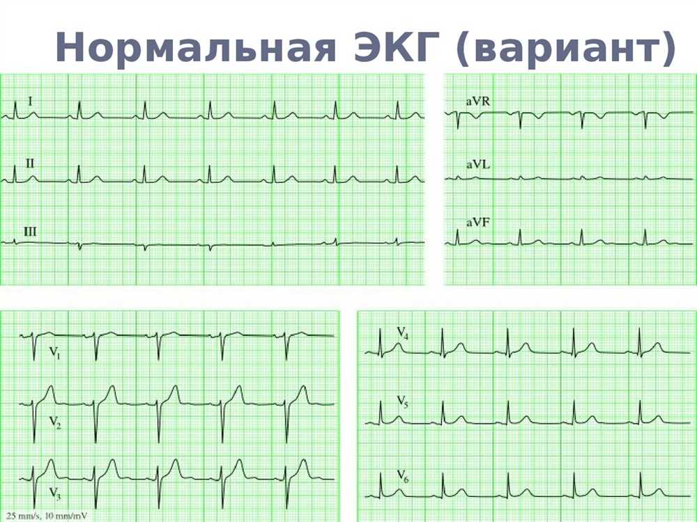
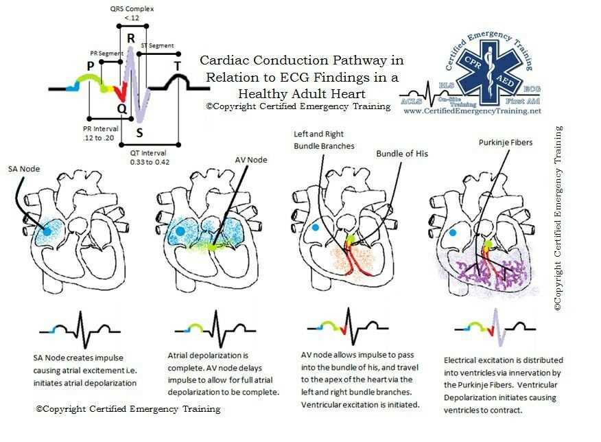
The electrical impulses recorded by the electrodes are represented as a graph on the EKG machine. The graph shows the different waves and intervals that are indicative of different aspects of heart function. The P wave represents the electrical impulse that causes the atria to contract, while the QRS complex represents the electrical impulse that causes the ventricles to contract. The T wave represents the recovery phase of the ventricles.
Interpreting the EKG
To interpret an EKG, healthcare professionals look at the various components of the graph, such as the shape, duration, and interval length. They analyze these components to identify any abnormalities and determine the overall health of the heart. For example, an enlarged QRS complex may indicate a blockage in the electrical pathway of the heart, while an irregular rhythm may suggest an arrhythmia.
By understanding the principles of an EKG and analyzing the different waves and intervals, healthcare professionals can gain valuable insights into a patient’s heart function and make appropriate diagnoses and treatment plans. This noninvasive test plays a crucial role in the detection and management of various heart conditions, ultimately helping to improve patient outcomes.
Importance of EKG in Cardiac Diagnosis
An electrocardiogram (EKG) is a vital tool in the diagnosis and management of cardiovascular conditions. It is a non-invasive test that records the electrical activity of the heart, providing valuable information about its rhythm and function. EKG readings can help healthcare professionals identify various heart conditions, assess the severity of the disease, and guide treatment decisions for patients.
One of the primary uses of EKG is in diagnosing arrhythmias, which are abnormal heart rhythms. By analyzing the patterns of electrical impulses displayed on the EKG, healthcare providers can identify irregularities that may be indicative of specific arrhythmias, such as atrial fibrillation or ventricular tachycardia. This information is crucial for determining the appropriate treatment approach, which may include medication, cardioversion, or even surgery.
Moreover, EKG can also assist in diagnosing acute myocardial infarction (heart attack). When a person experiences chest pain or other symptoms suggestive of a heart attack, an EKG can quickly assess the presence and location of myocardial ischemia (lack of blood flow to the heart muscle). The EKG can help identify ST-segment elevation, which is a strong indicator of an ongoing heart attack, prompting immediate intervention to restore blood flow and save the patient’s life.
Furthermore, EKG can be useful in monitoring patients with existing heart conditions, such as heart failure or coronary artery disease. Changes in the EKG readings over time can indicate worsening of the condition or the effectiveness of the prescribed treatment. Regular EKG monitoring can help healthcare providers adjust medication dosages, recommend lifestyle modifications, or consider additional interventions to optimize patient outcomes.
In summary, EKG plays a crucial role in the diagnosis and management of cardiac conditions. Its ability to assess heart rhythm, detect arrhythmias, diagnose myocardial infarction, and monitor ongoing heart conditions makes it an invaluable tool in the cardiovascular healthcare setting. Healthcare professionals rely on accurate EKG readings to make informed decisions and provide optimal care for patients with heart disease.
Steps to Conduct EKG Lab Quest 12
In order to conduct EKG Lab Quest 12, follow the steps below:
Step 1: Preparation
Ensure that all necessary equipment, including the EKG machine, electrodes, and recording paper, are properly set up and functioning correctly. Make sure you have the patient’s information, such as their name and date of birth, and explain the procedure to the patient to ensure their comfort and cooperation.
Step 2: Placement of Electrodes
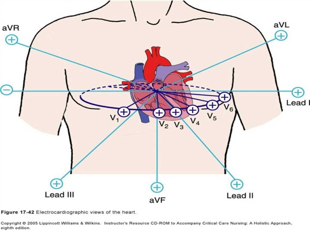
Apply the electrodes to the patient’s chest and limbs according to the standard lead placements. The right arm electrode should be placed on the wrist or upper forearm, the left arm electrode on the wrist or upper forearm, the right leg electrode on the ankle or lower leg, and the left leg electrode on the ankle or lower leg. The precordial (chest) electrodes should be placed in the specific locations indicated on the EKG machine.
Step 3: Recording the EKG
Start the EKG recording on the machine and ensure that the patient remains still during the recording to prevent interference. The machine will display and print the electrical activity of the patient’s heart, represented as a series of waves and intervals. Make sure to record the necessary duration of the EKG, as specified in the lab instructions.
Step 4: Interpretation and Analysis
Once the EKG recording is complete, analyze the waves and intervals to identify any abnormalities or irregularities in the heart’s electrical activity. Look for specific patterns or deviations from the normal EKG waveform, such as changes in the P wave, QRS complex, or T wave. Compare the EKG results to standard reference values to determine if any further evaluation or medical intervention is necessary.
Step 5: Documentation and Reporting
Document the findings of the EKG analysis and any relevant observations or notes. Use clear and concise language to describe any abnormalities or irregularities. Prepare a report summarizing the EKG results and any recommendations for further evaluation or treatment, if applicable.
By following these steps, you can successfully conduct EKG Lab Quest 12 and gather valuable information about the electrical activity of the heart.
Interpreting EKG Results from Lab Quest 12
The EKG, or electrocardiogram, is a valuable tool in analyzing the electrical activity of the heart. Lab Quest 12 provides a variety of EKG results that allow for detailed interpretation and analysis. By understanding the different components and measurements of an EKG, healthcare professionals can gather important information about a patient’s heart health.
One key aspect of interpreting EKG results is analyzing the P wave. The P wave represents the electrical activity of the atria, or the upper chambers of the heart. Its duration and shape can provide information about the conduction pathway through the atria and can help identify abnormalities such as atrial enlargement or a conduction delay.
The next element to consider is the QRS complex, which represents the electrical activity of the ventricles, or the lower chambers of the heart. The duration and amplitude of the QRS complex can indicate the overall health and integrity of the ventricles. Abnormalities in the QRS complex can indicate conditions such as ventricular hypertrophy or conduction abnormalities.
In addition to examining specific components, it is also important to analyze the overall rhythm of the EKG. The regularity of the heart rate, the presence of abnormal beats, and the presence of pauses or irregularities can all be indicative of underlying heart conditions. For example, an irregular rhythm may suggest atrial fibrillation, while a slow heart rate could indicate sinus bradycardia.
Overall, interpreting EKG results from Lab Quest 12 requires a thorough understanding of the different components and measurements of the EKG, as well as the ability to recognize patterns and abnormalities. By carefully analyzing the P wave, QRS complex, and overall rhythm, healthcare professionals can gain valuable insights into a patient’s heart health and make informed decisions regarding treatment and management.
Analyzing the Heart’s Electrical Activity with Lab Quest 12
Lab Quest 12 is a powerful tool used in the analysis of the heart’s electrical activity. This device allows researchers and medical professionals to gain valuable insights into the functioning of the heart and diagnose potential abnormalities. By recording and analyzing the electrocardiogram (ECG), Lab Quest 12 provides a detailed picture of the heart’s electrical signals and helps in understanding various cardiac conditions.
One of the key features of Lab Quest 12 is its ability to measure and display the different components of the ECG waveform. This includes the P wave, QRS complex, and T wave, which are crucial in determining the normality of the heart’s electrical conduction system. By examining the shape, duration, and timing of these components, researchers can identify any irregularities or abnormalities that may indicate underlying heart conditions.
Lab Quest 12 also offers advanced analysis tools that allow for the calculation of various parameters related to the heart’s electrical activity. These parameters include heart rate, PR interval, QRS duration, and QT interval. By comparing these values to established norms, medical professionals can assess the overall health of the heart and detect any deviations that may require further investigation or treatment.
Furthermore, Lab Quest 12’s data visualization capabilities enable researchers to easily interpret and present their findings. With the help of graphs and charts, they can visualize the changes in the ECG waveform over time, identify patterns, and make informed decisions regarding patient care. This makes Lab Quest 12 an invaluable tool in the field of cardiology, providing both qualitative and quantitative information about the heart’s electrical activity.
In conclusion, Lab Quest 12 plays a crucial role in analyzing the heart’s electrical activity. By recording and analyzing the ECG waveform, measuring various parameters, and visualizing the data, this device helps researchers and medical professionals uncover valuable insights and diagnose cardiac conditions effectively.
Common EKG Lab Quest 12 Patterns and Their Significance
An electrocardiogram, or EKG, is a test that measures the electrical activity of the heart. EKGs are commonly used in medical settings to assess the health of a patient’s heart and diagnose any potential issues. EKG Lab Quest 12 focuses on analyzing various EKG patterns and understanding their significance.
1. Sinus Rhythm
The sinus rhythm is the normal electrical activity of the heart. It is characterized by a regular heartbeat and a consistent pattern on the EKG. This pattern indicates that the heart’s electrical signals are originating from the sinus node, which is the natural pacemaker of the heart. A sinus rhythm is considered a healthy and normal pattern.
2. Atrial Fibrillation
Atrial fibrillation is a common arrhythmia characterized by an irregular and rapid heartbeat. On the EKG, atrial fibrillation is represented by chaotic and irregular waves, with no distinct P waves. This pattern indicates that the atria of the heart are quivering rather than contracting properly. Atrial fibrillation can increase the risk of blood clots and other heart-related complications.
3. Ventricular Tachycardia
Ventricular tachycardia is a potentially life-threatening arrhythmia characterized by a rapid heart rate originating in the ventricles. On the EKG, ventricular tachycardia is represented by wide QRS complexes with a rate greater than 100 beats per minute. This pattern indicates a disruption in the electrical signals of the heart and can lead to inadequate blood flow and cardiac arrest if left untreated.
4. Ventricular Fibrillation
Ventricular fibrillation is the most serious form of arrhythmia and is considered a medical emergency. It is characterized by chaotic and disorganized electrical activity in the ventricles, resulting in an extremely rapid and irregular heartbeat. On the EKG, ventricular fibrillation appears as a chaotic and erratic pattern with no discernible QRS complexes. Immediate medical intervention, such as defibrillation, is required to restore a normal heart rhythm.
5. ST-Segment Elevation
ST-segment elevation is a pattern on the EKG that can indicate a heart attack (myocardial infarction). It is characterized by an upward shift of the ST segment from the baseline. This pattern suggests that blood flow to a certain part of the heart is blocked, and the affected heart muscle is not receiving enough oxygen. Prompt medical intervention, such as angioplasty or thrombolytic therapy, is crucial to restore blood flow and prevent further damage to the heart.
Being able to recognize and interpret these common EKG patterns is essential for healthcare professionals involved in diagnosing and treating heart conditions. These patterns provide valuable insights into the electrical activity of the heart and can help guide appropriate medical interventions and treatments.