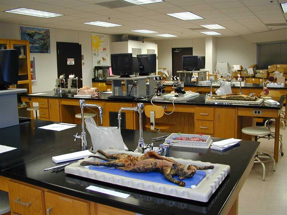
If you are currently studying anatomy and preparing for your first lab exam, you’re probably feeling a mixture of excitement and uncertainty. The lab exam can be challenging, but proper preparation and practice can significantly increase your chances of success. In this article, we will guide you through a comprehensive review of the topics that are likely to be covered in the anatomy lab exam 1, providing you with the knowledge and confidence you need to ace the test.
During the anatomy lab exam, you will be tested on your understanding of anatomical structures, their functions, and their relationships within the human body. This practical exam will require you to identify various structures, such as bones, muscles, organs, and blood vessels. To succeed, it is essential to develop a solid understanding of anatomical terminology, body regions, and the overall organization of the human body.
In addition to theoretical knowledge, the lab exam will also assess your ability to perform specific tasks related to anatomy. These may include the identification of structures on anatomical models, dissection of cadavers, or interpretation of medical imaging scans. To excel in these practical tasks, it is crucial to familiarize yourself with the anatomical landmarks, proper dissection techniques, and different imaging modalities commonly used in clinical practice.
Preparing for the anatomy lab exam can be an intensive process, but with the right mindset and study techniques, it can also be a rewarding one. Throughout this article, we will provide you with practice exercises, study tips, and valuable resources that will help you optimize your learning and achieve success in your anatomy lab exam 1.
Anatomy Lab Exam 1 Practice
In preparation for the upcoming Anatomy Lab Exam 1, it is important to engage in regular practice. This will help reinforce your understanding of the various anatomical structures and their functions. In this practice session, we will focus on identifying and labeling key structures of the human body.
One of the main areas of focus for Exam 1 is the skeletal system. You will be expected to identify and locate the major bones of the body, including the skull, spine, ribs, and limbs. It is important to familiarize yourself with the specific names and landmarks of these bones, as well as their functions within the body.
- Skull: The skull is composed of various bones, including the cranium and the facial bones. It serves to protect the brain, as well as house the structures involved in vision, hearing, and smell.
- Spine: The spine, also known as the vertebral column or backbone, is made up of individual vertebrae. It provides support for the body and protects the spinal cord, which transmits signals between the brain and the rest of the body.
- Ribs: The ribs are long, curved bones that form the rib cage. They protect the chest cavity and vital organs such as the heart and lungs.
- Limbs: The limbs consist of the upper limbs (arms) and lower limbs (legs). They allow for movement and manipulation of objects, as well as provide support for the body.
During the Anatomy Lab Exam 1, you will be presented with various specimens, models, and visuals to identify and label. By regularly practicing in the lab and reviewing the relevant anatomical structures, you will be well-prepared for this exam. Remember to use anatomical terminology and proper labeling techniques to ensure accurate identification of structures.
Overview of the Human Body
The human body is a complex and intricate system that is made up of different organs, tissues, and cells. These components work together to maintain the body’s overall health and functionality. Understanding the structure and function of the human body is essential for healthcare professionals, as it forms the basis of medical knowledge and practice.
Anatomy is the study of the structure of the human body. It involves the examination and identification of the different organs, tissues, and structures that make up the body. This detailed understanding of anatomy allows healthcare professionals to accurately diagnose and treat various medical conditions.
Physiology is the study of how the different parts of the body function and work together. It examines the processes and mechanisms that occur within the body, such as digestion, respiration, and circulation. By studying physiology, healthcare professionals can gain insights into how these processes are regulated and maintained.
The human body can be divided into several major systems, each with its own specific functions and structures. These include the skeletal system, muscular system, cardiovascular system, respiratory system, digestive system, nervous system, and reproductive system. Each of these systems plays a vital role in maintaining the body’s overall health and functionality.
- The skeletal system provides support and structure to the body, and is composed of bones, ligaments, and cartilage.
- The muscular system enables movement and is responsible for generating force and heat.
- The cardiovascular system transports oxygen, nutrients, hormones, and waste products throughout the body.
- The respiratory system facilitates the exchange of oxygen and carbon dioxide, allowing for respiration.
- The digestive system breaks down food into nutrients that can be absorbed and utilized by the body.
- The nervous system controls and coordinates bodily functions through electrochemical signals.
- The reproductive system is responsible for the production of offspring.
Overall, the human body is a complex and integrated system that requires the proper functioning of all its parts for optimal health and well-being.
The Skeletal System
The skeletal system is an essential component of the human body, providing structural support, protection for vital organs, and the ability to move. It is composed of bones, joints, cartilage, and other connective tissues that work together to form the framework of the body.
There are 206 bones in the adult human body, each with a specific shape, size, and function. These bones can be divided into two main categories: axial skeleton and appendicular skeleton. The axial skeleton includes the bones that make up the skull, vertebral column, and ribcage, while the appendicular skeleton consists of the bones of the limbs, pelvis, and shoulder girdle.
The skeletal system serves multiple important functions. Firstly, it provides support and structure, allowing the body to maintain its shape and integrity. The bones act as a strong scaffold for the muscles, tendons, and ligaments that enable movement and stability. Additionally, the skeletal system protects vital organs such as the brain, heart, and lungs. For example, the skull protects the brain, while the ribcage shields the heart and lungs from injury.
Moreover, the skeletal system plays a crucial role in the production and storage of blood cells. In the bone marrow, red and white blood cells are produced, which are essential for oxygen transport and immune system function. Additionally, certain bones, such as the femur and humerus, serve as storage sites for important minerals like calcium and phosphorus, which are critical for maintaining healthy bones and overall bodily functions.
In conclusion, the skeletal system is an intricate network of bones, joints, and connective tissues that provides support, protection, and mobility to the human body. Understanding the structure and function of the skeletal system is crucial for medical professionals, as it forms the foundation for understanding bodily movements, injuries, and diseases.
The Muscular System
The muscular system is a complex network of muscles that enable movement, provide support to the body, and facilitate various bodily functions. Muscles are essential for locomotion, maintaining posture, and generating heat to regulate body temperature.
There are three types of muscles in the muscular system: skeletal muscles, smooth muscles, and cardiac muscles. Skeletal muscles, also known as voluntary muscles, are attached to bones and are responsible for voluntary movements such as walking, running, and lifting weights. Smooth muscles are found in the walls of organs and blood vessels, and are responsible for involuntary movements such as peristalsis. Cardiac muscles are found in the heart and are responsible for the contraction and relaxation of the heart, ensuring the continuous pumping of blood throughout the body.
Each muscle is composed of individual muscle fibers, which are long, slender cells that are capable of contracting and relaxing. These muscle fibers are bundled together in fascicles, which are surrounded by connective tissue called perimysium. The connective tissue not only provides support and protection to the muscle fibers, but also allows them to work together efficiently.
During muscle contraction, actin and myosin, two types of proteins, interact to shorten the muscle fibers. This generates a pulling force on the bones and creates movement. The nervous system plays a crucial role in muscle contraction by sending signals to the muscles to initiate and control movement.
In summary, the muscular system is a complex network of muscles that enable movement, support the body, and facilitate various bodily functions. Understanding the structure and function of muscles is essential for studying human anatomy and physiology.
The Cardiovascular System
The cardiovascular system, also known as the circulatory system, is responsible for the transport of blood, oxygen, nutrients, hormones, and other essential substances throughout the body. It is composed of the heart, blood vessels, and blood.
The heart acts as a pump that propels the blood through the blood vessels. It is a muscular organ made up of four chambers – two atria and two ventricles. The right side of the heart receives deoxygenated blood from the body and pumps it to the lungs for oxygenation, while the left side receives oxygenated blood from the lungs and pumps it to the rest of the body.
The blood vessels include arteries, veins, and capillaries. Arteries carry oxygenated blood away from the heart to the various tissues and organs of the body, while veins carry deoxygenated blood back to the heart. Capillaries are tiny blood vessels that allow for the exchange of oxygen, nutrients, and waste products between the blood and surrounding tissues.
The blood is a specialized connective tissue that consists of red blood cells, white blood cells, platelets, and plasma. Red blood cells are responsible for carrying oxygen to the tissues and removing carbon dioxide, while white blood cells play a crucial role in the body’s immune response. Platelets are vital for blood clotting, and plasma carries nutrients, hormones, and waste products.
- In summary, the cardiovascular system plays a crucial role in maintaining the body’s overall function. It ensures that all tissues receive the necessary oxygen and nutrients while removing waste products.
- The heart, blood vessels, and blood work together to achieve this function, allowing for proper circulation throughout the body.
- Any disruption or malfunction in the cardiovascular system can lead to various diseases and conditions, such as heart disease, high blood pressure, or stroke.
Understanding the anatomy and function of the cardiovascular system is essential for healthcare professionals, as it helps in diagnosing and treating cardiovascular-related disorders.
The Respiratory System

The respiratory system is a crucial part of the human body, responsible for the exchange of oxygen and carbon dioxide. Composed of various organs, including the nose, throat, trachea, lungs, and diaphragm, this system works together to facilitate respiration and maintain homeostasis. Let’s take a closer look at the components and functions of the respiratory system.
Organs of the Respiratory System
The respiratory system consists of several organs that work in harmony to perform vital functions. The pathway of air begins in the nose, where incoming air is filtered, warmed, and humidified. From there, the air passes through the throat, or pharynx, and then enters the trachea, or windpipe. The trachea branches into two bronchi, which further divide into smaller bronchioles. Finally, the bronchioles lead to the alveoli, the tiny air sacs in the lungs where gas exchange occurs.
The lungs are the primary organs of the respiratory system. Protected by the ribcage, these spongy, elastic structures are responsible for inhaling oxygen and exhaling carbon dioxide. Each lung is divided into lobes – the right lung has three lobes, while the left lung has two. Together, the lungs provide a large surface area for gas exchange, ensuring the body receives enough oxygen and eliminates metabolic waste.
Functions of the Respiratory System

The primary function of the respiratory system is to supply the body with oxygen and remove carbon dioxide, maintaining optimal levels of blood gases. Inhalation, or inspiration, occurs when the diaphragm contracts and the rib muscles expand, increasing the volume of the thoracic cavity. This creates a pressure difference, causing air to rush into the lungs. Exhalation, or expiration, happens when the diaphragm relaxes and the rib muscles contract, reducing the volume of the thoracic cavity and forcing air out of the lungs.
In addition to gas exchange, the respiratory system also plays a role in vocalization, as air passing through the vocal cords produces sound. It is also involved in regulating body temperature, as breathing helps dissipate heat through exhaled air. Lastly, the respiratory system participates in the sense of smell, with odorous molecules being detected by specialized cells in the nasal cavity.
In conclusion, the respiratory system is essential for the survival and functioning of the human body. It enables the exchange of oxygen and carbon dioxide, supports speech and vocalization, helps regulate body temperature, and contributes to the sense of smell. Understanding the anatomy and physiology of the respiratory system is crucial for healthcare professionals and individuals interested in maintaining optimal respiratory health.