
If you’re studying anatomy and trying to learn about bones, one great way to reinforce your knowledge is through coloring. Coloring pages can help you visualize the different parts of a bone and understand their functions. In this article, we will provide answers to some common questions about the anatomy of a bone coloring pages.
Firstly, let’s talk about the basic structure of a bone. Bones are made up of different layers, including the periosteum, compact bone, spongy bone, and bone marrow. The periosteum is the outermost layer of the bone, providing protection and serving as a site for muscle attachment. The compact bone is the strong, dense layer that makes up most of the bone’s mass, providing support and strength. The spongy bone, also known as cancellous bone, consists of a network of trabeculae and is responsible for storing bone marrow. Lastly, the bone marrow is a soft, fatty tissue found inside the bone and is responsible for producing blood cells.
When coloring a bone, it’s important to understand the different parts that make up its anatomy. Common structures to color include the diaphysis, epiphysis, metaphysis, and articular cartilage. The diaphysis is the long shaft of the bone, while the epiphysis refers to the ends of the bone. The metaphysis is the area where the diaphysis and epiphysis meet, and is responsible for bone growth. The articular cartilage, on the other hand, covers the ends of the bones where they meet to form joints, providing cushioning and reducing friction.
In conclusion, coloring pages can be a helpful tool for learning about the anatomy of a bone. By understanding the different layers and structures that make up a bone, you can enhance your knowledge and retain information more effectively. So grab your coloring pencils and start exploring the fascinating world of bone anatomy!
Anatomy of a Bone Coloring Answers
Understanding the anatomy of a bone is crucial for students studying biology or anatomy. Coloring diagrams of bones can help reinforce knowledge of bone structure and function. Here are some answers to commonly colored sections of a bone:
Cortex:
The cortex is the hard, dense outer layer of a bone. It provides strength and support to the bone. When coloring the cortex, it is often shaded in a darker color to represent its compact nature.
Medullary Cavity:
The medullary cavity is the central cavity within the bone. It is filled with yellow marrow, which is responsible for storing fat. When coloring the medullary cavity, it is typically left blank or shaded in a light color to represent its hollow nature.
Periosteum:
The periosteum is the tough, fibrous membrane that covers the outside of the bone. It is important for bone growth and repair. When coloring the periosteum, it is often shaded in a lighter color to differentiate it from the cortex.
Epiphysis:
The epiphysis is the ends of the bone. It is covered in a layer of cartilage and plays a role in joint movement. When coloring the epiphysis, it can be shaded in a different color to highlight its unique structure.
Coloring a bone diagram can be a fun and interactive way to learn about the anatomy of a bone. By coloring different sections, students can visually reinforce their understanding of bone structure and function.
Types of Bones
In the human body, there are five main types of bones: long bones, short bones, flat bones, irregular bones, and sesamoid bones.
Long Bones
Long bones are characterized by their elongated shape and include bones such as the femur (thigh bone) and humerus (upper arm bone). They provide support and facilitate movement by acting as levers for the muscles. Long bones consist of a hard outer layer of compact bone that encloses a spongy inner layer called the marrow cavity, which contains bone marrow.
Short Bones
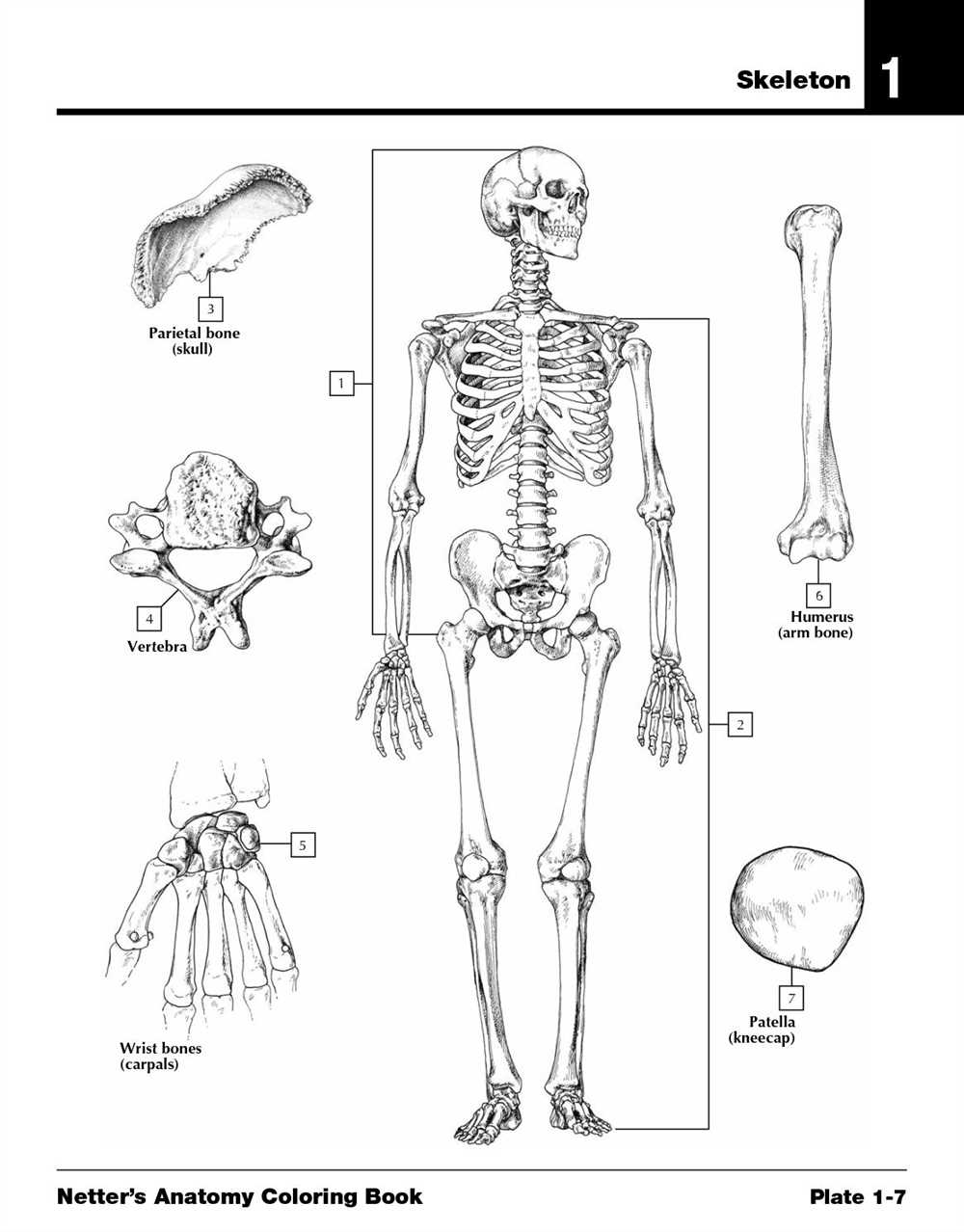
Short bones are typically cube-shaped and provide stability and support. Examples of short bones include the bones of the wrist (carpals) and ankle (tarsals). Despite their small size, short bones play an important role in weight-bearing and joint movement.
Flat Bones
Flat bones have a flattened, thin shape and provide protection for internal organs. Examples of flat bones include the skull, ribs, and scapulae. They consist of two layers of compact bone with a layer of spongy bone in between.
Irregular Bones
Irregular bones have a unique shape that does not fit into the other categories. They provide support and protection to vital organs and help with various bodily functions. Examples of irregular bones include the vertebrae, facial bones, and pelvic bones.
Sesamoid Bones
Sesamoid bones are small, round bones that develop within tendons, typically in response to strain or stress. The patella (kneecap) is an example of a sesamoid bone. These bones help to reduce friction and increase the mechanical advantage of the muscles.
In summary, understanding the different types of bones in the human body is essential for comprehending their functions and roles in supporting the body’s structure and facilitating movement.
Structure of a Bone
A bone is a complex structure, made up of several different components, all working together to provide strength, support, and protection to the body. The main components of a bone include the periosteum, compact bone, spongy bone, and bone marrow.
Periosteum: The periosteum is a tough, fibrous membrane that covers the outer surface of the bone. It provides nutrients to the bone and plays a crucial role in the repair and growth of the bone.
Compact Bone: The compact bone is the dense outer layer of the bone. It is made up of tightly packed osteons, or structural units, that give the bone its strength and rigidity. The compact bone also contains small channels called canaliculi, which allow for the exchange of nutrients and waste between the bone cells.
Spongy Bone: The spongy bone, also known as trabecular or cancellous bone, is found on the inside of the bone, beneath the compact bone. It consists of a network of bony struts called trabeculae, which provide structural support while also allowing for the passage of blood vessels and bone marrow.
Bone Marrow: The bone marrow is a soft, gelatinous tissue found within the cavities of certain bones. It is responsible for the production of different types of blood cells, including red blood cells, white blood cells, and platelets. There are two types of bone marrow: red marrow, which is involved in blood cell production, and yellow marrow, which consists mainly of fat cells and serves as a storage site for energy.
In summary, the structure of a bone is a complex and intricate system that includes the periosteum, compact bone, spongy bone, and bone marrow. Each component has its own unique function and together they work to ensure the strength, support, and functionality of the bone.
Functions of Bones
Bones are an essential component of the skeletal system and serve several important functions in the human body.
Support and Protection:
One of the primary functions of bones is to provide support and protection for the body. Bones form the structural framework that supports our muscles, organs, and tissues. They give our body shape and provide a solid foundation for our movements. Additionally, bones protect delicate internal organs such as the brain, heart, and lungs from any external impact or injury.
Movement:
Bones also play a crucial role in enabling movement. They serve as an attachment point for muscles, ligaments, and tendons, which work together to facilitate motion. When muscles contract, they pull on the bones, causing them to move. This allows us to perform various activities such as walking, running, and lifting objects.
Blood Cell Production:
The bones are responsible for the production of blood cells, a process known as hematopoiesis. The bone marrow, found within the spongy part of bones, produces red blood cells, white blood cells, and platelets. These blood cells are vital for carrying oxygen, fighting infections, and clotting blood.
Mineral Storage:
Bones act as a reservoir for minerals, primarily calcium and phosphorus. These minerals are essential for maintaining proper bone density and strength. When the body needs more minerals, such as during pregnancy or in case of a deficiency, it can draw from the bones to meet the demand. Conversely, when there is an excess of minerals, the bones can store them for future use.
Endocrine Regulation:
Bones also play a role in endocrine regulation. They produce hormones such as osteocalcin, which helps regulate glucose metabolism and insulin secretion. Osteocalcin also influences fertility, energy expenditure, and cognition.
In conclusion, bones have multiple functions that are vital for the overall health and functioning of the human body. They provide support and protection, enable movement, produce blood cells, store minerals, and participate in endocrine regulation. Maintaining healthy bones is essential for a healthy and active lifestyle.
Bone Formation and Growth
Bone formation, also known as osteogenesis, is a complex process that occurs throughout life to ensure the growth, maintenance, and repair of the skeletal system. It involves the synthesis and mineralization of bone tissue, as well as the remodeling and resorption of old or damaged bone. The cells responsible for bone formation are known as osteoblasts, which are derived from mesenchymal stem cells.
During embryonic development, bones are initially formed through a process called endochondral ossification, where a cartilage model is gradually replaced by bone tissue. This process begins with the differentiation of mesenchymal cells into chondroblasts, which form a cartilage model that serves as a template for bone formation. Osteoblasts then invade the cartilage model and deposit osteoid, a collagen-rich matrix that eventually mineralizes to form bone.
In addition to endochondral ossification, bones can also be formed through intramembranous ossification. This process occurs primarily in flat bones, such as the skull and clavicles. It involves the direct differentiation of mesenchymal cells into osteoblasts, which then secrete osteoid and mineralize it to form bone. Intramembranous ossification is responsible for the growth and shaping of these bones during development.
Throughout life, bone growth occurs through a process called bone remodeling. This process involves the coordinated activity of osteoblasts and osteoclasts, a type of bone-resorbing cell. Osteoblasts deposit new bone on the surface of existing bone, while osteoclasts resorb and remove old or damaged bone tissue. The balance between these two processes ensures that the skeleton remains strong and healthy.
In conclusion, bone formation and growth are essential processes for the development and maintenance of the skeletal system. These processes involve the differentiation and activity of specialized cells, such as osteoblasts and osteoclasts. Understanding the mechanisms of bone formation and growth can help in the diagnosis and treatment of various bone disorders and injuries.
Bone Coloring Process
The process of coloring a bone involves several steps to ensure a realistic and accurate representation of its anatomy. This process is commonly used in educational settings, such as medical schools or anatomy classes, to help students understand the intricate structure of bones in the human body.
1. Preparation: Before starting the coloring process, it is essential to properly clean and dry the bone to remove any dirt, oils, or other contaminants. This step ensures that the coloring materials adhere well to the bone’s surface.
2. Selection of Coloring Materials: There are various coloring materials available for bone coloring, such as acrylic paints, colored pencils, or specialized bone coloring kits. The choice of coloring material depends on the desired level of detail and the intended use of the colored bone.
3. Color Mapping: Color mapping is an important step in the bone coloring process. It involves identifying and mapping the different anatomical structures of the bone, such as the compact bone, spongy bone, periosteum, and various types of bone marrow. Each structure is assigned a specific color or shade to differentiate it from others.
4. Application of Colors: Once the color mapping is complete, the chosen coloring materials are applied to the bone’s surface. Depending on the material used, this may involve brushing, drawing, or airbrushing the colors onto the bone. Care must be taken to ensure even coverage and precise application to accurately represent the anatomy.
5. Finishing Touches: After the colors have been applied, additional details and textures can be added to enhance the realism of the colored bone. This may include adding shading, highlights, or fine lines to represent blood vessels or muscle attachments. Varnishing or sealing the colored bone may also be done to protect the colors and improve longevity.
Overall, the bone coloring process is an intricate and meticulous task that requires attention to detail and knowledge of bone anatomy. Through this process, students can gain a deeper understanding of the structures within bones and visualize their organization in a three-dimensional form. This hands-on approach to learning can significantly enhance the educational experience and aid in the retention of anatomical knowledge.
Common Bone Disorders
Bones are essential for the structure and support of the human body. However, they can be affected by various disorders that can have a significant impact on a person’s quality of life. Some of the most common bone disorders include:
Osteoporosis: Osteoporosis is a condition characterized by low bone density and weakened bones. It occurs when the body loses too much bone mass or doesn’t produce enough new bone. This can increase the risk of fractures and make bones more susceptible to injuries.
Osteoarthritis: Osteoarthritis is a degenerative joint disorder that often affects the bones in the hands, knees, hips, and spine. It occurs when the protective cartilage that cushions the ends of bones wears down over time, resulting in pain, stiffness, and reduced mobility.
Rheumatoid arthritis: Rheumatoid arthritis is an autoimmune disorder that primarily affects the joints, but it can also cause inflammation in the bones. This chronic condition leads to painful and swollen joints, as well as bone erosion and deformities in severe cases.
Fractures: Fractures, or broken bones, can occur due to traumatic injuries or as a result of bone weakness. Common types of fractures include greenstick fractures, spiral fractures, and comminuted fractures. Treatment for fractures typically involves immobilization and, in some cases, surgical intervention.
Osteomyelitis: Osteomyelitis is a bone infection that can be caused by a variety of bacteria. It can occur as a result of an open fracture, surgical procedure, or bloodstream infection. This condition can lead to severe pain, swelling, and bone damage if left untreated.
Bone cancer: Bone cancer can affect any bone in the body and can occur as a primary cancer or as a metastasis from another part of the body. Common types of bone cancer include osteosarcoma, chondrosarcoma, and Ewing sarcoma. Treatment options for bone cancer may include surgery, radiation therapy, and chemotherapy.
These are just a few examples of the many bone disorders that can affect individuals. It is important to seek medical attention if you experience any symptoms or have concerns about your bone health to receive an accurate diagnosis and appropriate treatment.