
Gel electrophoresis is a widely used technique in molecular biology and genetics that allows scientists to separate and analyze DNA fragments or proteins based on their size and charge. This powerful tool has revolutionized the field, enabling researchers to gain valuable insights into various biological processes and phenomena.
In a gel electrophoresis virtual lab, students are provided with a simulated environment where they can perform experiments and explore the principles of this technique. By answering specific questions related to their virtual experiments, students can deepen their understanding of electrophoresis and its applications in real research settings.
Through the virtual lab, students can learn about the different components and processes involved in gel electrophoresis, such as the gel matrix, buffer solution, and the application of an electric field. They can also understand how charged molecules move through the gel matrix based on their charge and size, leading to the separation and visualization of bands on the gel.
Additionally, the virtual lab allows students to investigate the factors that affect the migration of molecules in gel electrophoresis, such as the concentration of the gel, voltage, and the size of the molecules being analyzed. By manipulating these variables, students can observe the impact on the separation and analysis of DNA or proteins, deepening their understanding of the technique’s principles.
Gel Electrophoresis Virtual Lab Answers: A Comprehensive Guide
Gel electrophoresis is a widely used technique in molecular biology to separate and analyze DNA, RNA, or protein fragments based on their size and charge. In a traditional laboratory setting, gel electrophoresis involves running a gel containing a matrix, usually agarose or polyacrylamide, with an electric current applied to it. However, with the advancement of technology, virtual labs have become popular, allowing scientists and students to simulate gel electrophoresis experiments.
Virtual labs provide a convenient and cost-effective alternative to traditional lab experiments. They offer an interactive platform where users can manipulate various experimental parameters, such as gel concentration, voltage, and sample size, to observe how these factors affect the separation of DNA fragments. In addition, virtual labs often include pre-programmed scenarios with different DNA samples that users can analyze and interpret.
To get the most accurate and reliable results from a gel electrophoresis virtual lab, it is important to consider some key factors. Firstly, it is crucial to understand the principles behind gel electrophoresis and how the technique separates DNA fragments based on their size and charge. This knowledge will help in interpreting the results obtained from the virtual lab.
Secondly, it is essential to carefully follow the instructions provided in the virtual lab, including setting the appropriate gel concentration and running the gel at the recommended voltage and time. These parameters are critical for obtaining optimal separation of DNA fragments.
Lastly, when analyzing the results, it is important to consider the positions and sizes of the DNA fragments observed on the virtual gel. By comparing the band positions and sizes to a molecular weight ladder or a known DNA sample, users can estimate the size of their DNA fragments.
Overall, a gel electrophoresis virtual lab provides an engaging and educational experience for users to learn and practice the principles of gel electrophoresis. By following the principles of the technique and carefully analyzing the results, users can obtain valuable insights into the separation of DNA fragments and develop a better understanding of molecular biology.
Exploring the Concept of Gel Electrophoresis
Gel electrophoresis is a powerful technique used in molecular biology to separate and analyze DNA, RNA, and proteins. This method allows researchers to determine the size and charge of biomolecules, as well as study their interactions and functions. Gel electrophoresis plays a crucial role in many areas of scientific research, including genetics, forensics, and biotechnology.
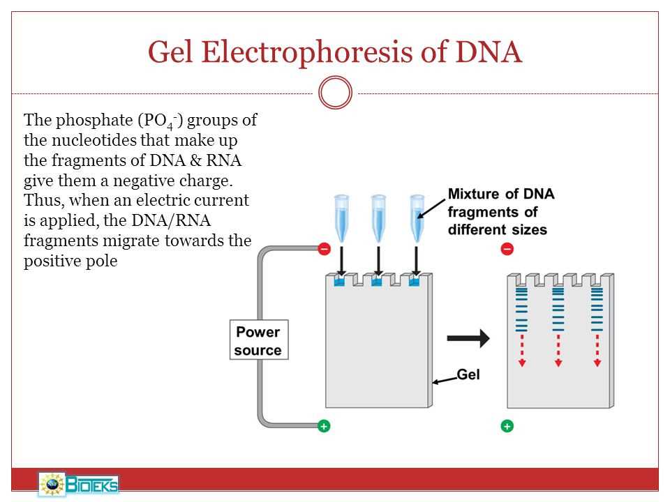
During gel electrophoresis, an electric current is applied to a gel matrix containing the biomolecules of interest. Because DNA, RNA, and proteins have negative charges, they are attracted to the positive electrode and move through the gel. The smaller molecules move more quickly through the gel, while the larger ones move slower. This separation is achieved due to the size and charge differences among the biomolecules.
The gel matrix used in gel electrophoresis can be made of different materials, such as agarose or polyacrylamide. Agarose gel electrophoresis is commonly used for separating DNA molecules, while polyacrylamide gel electrophoresis is used for separating proteins and smaller DNA fragments.
Once the biomolecules have migrated through the gel, they can be visualized using various staining methods. For DNA and RNA, fluorescent dyes such as ethidium bromide or SYBR Green are commonly used. For proteins, staining with Coomassie Brilliant Blue or silver stain is frequently employed. The stained gels can then be analyzed and documented using imaging systems or densitometers. After gel electrophoresis, the separated biomolecules can be extracted from the gel for further downstream applications, such as sequencing or cloning.
In conclusion, gel electrophoresis is a versatile and fundamental technique in molecular biology. It allows researchers to separate, analyze, and visualize DNA, RNA, and proteins based on their size and charge. This technique has revolutionized various fields of scientific research and is continuously improving with the development of new technologies and methods.
Understanding the Purpose of Gel Electrophoresis
Gel electrophoresis is a widely used technique in molecular biology and genetics that allows scientists to separate and analyze DNA, RNA, and proteins based on their size and charge. This technique plays a crucial role in various areas of research, such as genetic mapping, DNA sequencing, and forensic analysis.
The main purpose of gel electrophoresis is to separate DNA, RNA, or proteins into distinct bands or bands according to their molecular weight. The process involves applying an electric current to a gel matrix, usually made of agarose or polyacrylamide, which creates a charge gradient. Depending on their size and charge, molecules will migrate through the gel at different speeds, resulting in their separation and formation of distinct bands in the gel.
The separated molecules can then be visualized using staining techniques, such as ethidium bromide or fluorescent dyes, which bind to the nucleic acids and make them visible under UV light. The intensity and size of the bands can provide valuable information about the molecular weight, quantity, and purity of the molecules being analyzed, allowing researchers to make important conclusions about the sample.
Gel electrophoresis is an essential tool in genetic research because it allows scientists to identify genetic variations, such as mutations or polymorphisms, and compare different samples. It can also be used to determine the size of DNA or RNA fragments, which is crucial for applications like cloning or gene expression analysis. Overall, gel electrophoresis is a powerful technique that has revolutionized the field of molecular biology and provided valuable insights into the structure and function of biological molecules.
Step-by-Step Procedure of the Gel Electrophoresis Virtual Lab
In this virtual lab, we will be conducting gel electrophoresis, a technique used in molecular biology to separate and analyze DNA fragments based on their size. Gel electrophoresis is an essential tool in genetic research and forensics, allowing scientists to identify and compare DNA samples.
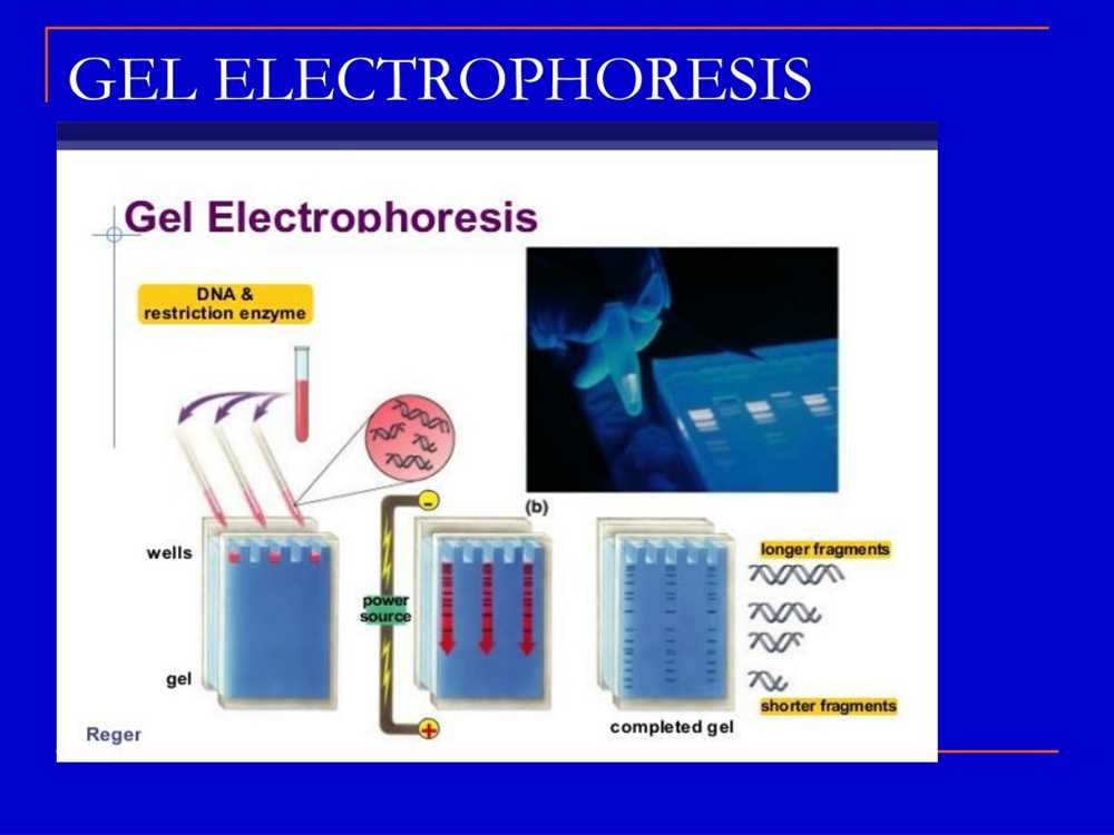
1. Set up the experiment:
Start by opening the virtual lab software and selecting the gel electrophoresis experiment. Make sure you have all the necessary materials, including a gel, DNA samples, and a DNA ladder. Place the gel in the electrophoresis chamber and add a buffer solution to create an even electric field.
2. Load the DNA samples:
Using micropipettes, carefully load the DNA samples onto the wells of the gel. The samples should be placed in separate wells, and the DNA ladder should be loaded in a separate well as a size reference. Make sure to record the positions of the DNA samples and ladder in the virtual lab software.
3. Run the electrophoresis:
Once the DNA samples are loaded, close the electrophoresis chamber and start the run. The DNA fragments will migrate through the gel towards the positive electrode, with smaller fragments moving faster and traveling further. Allow the electrophoresis to run for the specified time in the virtual lab.
4. Analyze the results:
After the electrophoresis run is complete, the DNA fragments will be separated based on their size. Use the virtual lab software to examine and analyze the gel image. You can measure the distance traveled by each DNA fragment and calculate their sizes using the size reference provided by the DNA ladder. Compare the patterns and sizes of the DNA fragments in different samples to draw conclusions about their genetic makeup.
5. Interpret the data:
Based on the analysis of the gel image, interpret the results of the gel electrophoresis. Look for similarities and differences in the DNA fragment patterns of the samples. Determine the sizes of unknown DNA fragments by comparing them to the DNA ladder. This information can help answer research questions or provide valuable insights in genetic studies and forensic investigations.
Analyzing Results and Interpretation of Gel Electrophoresis
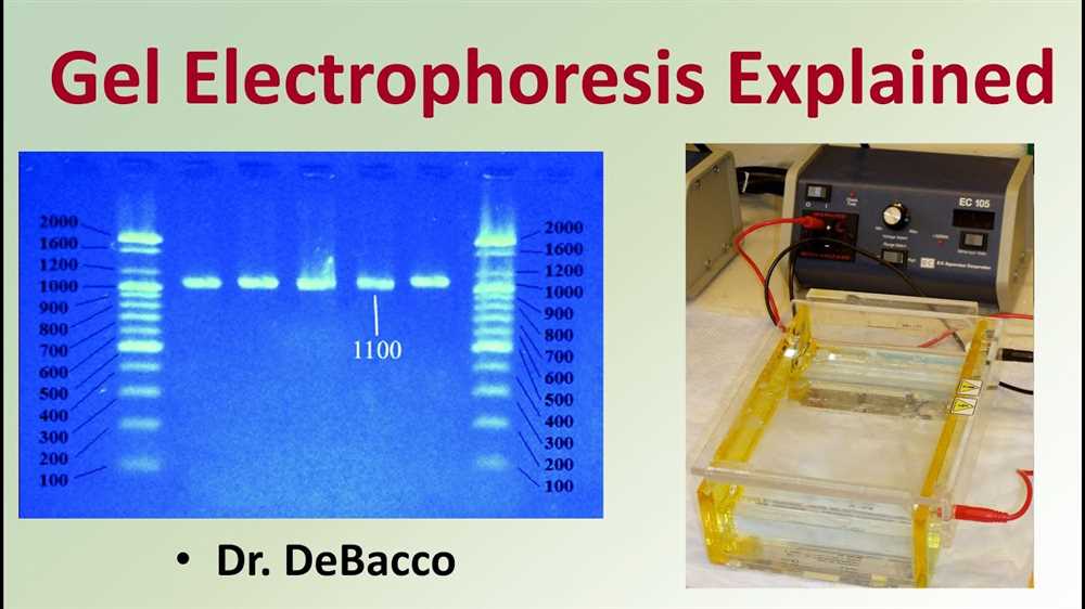
After running a gel electrophoresis experiment, it is necessary to analyze and interpret the results in order to understand the genetic information that has been separated. The first step in analyzing the results is to examine the gel itself. The gel electrophoresis creates distinct bands of different sizes, with the smaller fragments migrating further along the gel than the larger fragments.
The next step is to compare the results to a known DNA ladder, also known as a marker or standard. The DNA ladder contains DNA fragments of known sizes, and by comparing the bands on the gel to the ladder, it is possible to estimate the size of the fragments in the sample. This allows researchers to identify any genetic mutations or variations present.
In addition to estimating fragment size, gel electrophoresis can also provide information about the quantity of DNA present. The intensity of the bands on the gel can be used as a rough estimate of the amount of DNA, with darker and more intense bands indicating higher concentrations.
Overall, gel electrophoresis is a powerful tool for analyzing genetic information. By carefully examining the bands on the gel and comparing them to known standards, researchers can gain valuable insights into the size, quantity, and variations in DNA fragments. This information is crucial for understanding genetic diseases, conducting forensic analysis, and conducting research in genetics and molecular biology.
Common Challenges and Troubleshooting Tips
Working with gel electrophoresis can sometimes present challenges that can affect the results of your experiment. Here are some common issues and troubleshooting tips to help you overcome them:
1. Uneven or smeared bands on the gel:
- Possible cause: Uneven loading of the samples or incorrect application of the loading dye.
- Troubleshooting tip: Ensure that the samples are loaded evenly and apply the loading dye carefully to ensure consistent results.
2. No bands or faint bands on the gel:
- Possible cause: Insufficient or degraded DNA in the samples.
- Troubleshooting tip: Check the quality and concentration of your DNA samples. Use fresh samples or consider using a DNA extraction kit to obtain higher-quality DNA.
3. High background noise or smearing throughout the gel:
- Possible cause: Contamination of the gel or buffer.
- Troubleshooting tip: Ensure that all equipment and materials used are clean and free from contamination. Use fresh gel and buffer solutions to minimize background noise.
4. Overloading of the gel:
- Possible cause: Too much DNA loaded onto the gel, causing overlapping bands or smearing.
- Troubleshooting tip: Optimize the amount of DNA loaded onto the gel. If necessary, dilute your samples to ensure better separation of bands.
By being aware of these common challenges and following the troubleshooting tips, you can enhance the quality and reliability of your gel electrophoresis results. Remember to carefully evaluate each step of the experimental procedure and make adjustments as needed to overcome any issues that may arise.
Applications of Gel Electrophoresis in Research and Medicine
Gel electrophoresis is a widely used technique in research and medicine for various applications. It allows scientists to separate and analyze different biological molecules based on their size and charge. This technique has revolutionized the fields of genetics, biochemistry, and molecular biology, contributing to important discoveries and advancements in these areas.
One of the primary applications of gel electrophoresis is in DNA analysis. By running a sample of DNA through a gel and subjecting it to an electric field, scientists can separate the DNA fragments based on their size. This technique is used in forensic science to identify suspects and determine relationships between individuals. Gel electrophoresis is also used in genetic research to study patterns of inheritance and identify genetic mutations associated with diseases.
In addition to DNA analysis, gel electrophoresis is also used for protein analysis. Proteins can be separated by their size, charge, or both using gel electrophoresis. This technique is often used to study protein expression levels, identify protein-protein interactions, and analyze post-translational modifications. It is an essential tool in the field of proteomics, which aims to study and understand the functions and interactions of all proteins in an organism.
Gel electrophoresis is also used in medical diagnostics. It can be used to analyze samples from patients to detect the presence of specific genetic mutations, infectious agents, or abnormal proteins. This information can aid in the diagnosis and prognosis of various diseases, such as genetic disorders, viral infections, and cancer. Gel electrophoresis is an important tool in personalized medicine, allowing healthcare providers to tailor treatment plans based on a patient’s specific genetic makeup.
In summary, gel electrophoresis has a wide range of applications in research and medicine. It is a versatile technique that allows for the separation and analysis of DNA and proteins, providing valuable insights into various biological processes and diseases. Its widespread use in these fields highlights its importance in advancing scientific knowledge and improving healthcare outcomes.