
Understanding human karyotypes is essential in studying genetics and identifying genetic abnormalities. A karyotype is a visual representation of an individual’s chromosomes, which are organized pairs of DNA molecules that carry genetic information. By analyzing the structure and number of chromosomes, scientists can determine if there are any abnormalities or genetic disorders present.
In this exercise, students are tasked with cutting out and arranging the chromosomes to create a karyotype. By doing so, they gain a hands-on understanding of how chromosomes are organized and how they can be used to identify genetic disorders. The exercise helps develop critical thinking skills and allows students to apply their knowledge of genetics in a practical way.
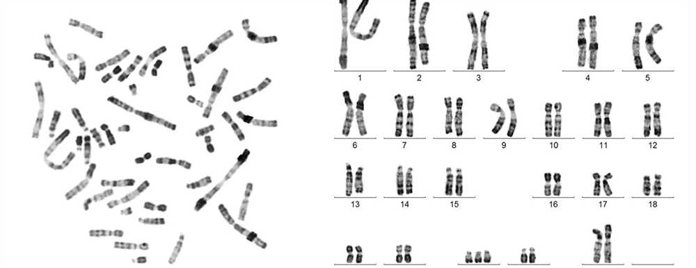
The cut and paste answers for the human karyotype exercise involve arranging the chromosomes in pairs according to their size and visual characteristics. By doing this, students can create a karyotype that accurately represents a specific individual’s genetic makeup. The exercise may also include identifying any abnormalities or genetic disorders based on the arrangement and structure of the chromosomes.
What is a human karyotype?
A human karyotype refers to the arrangement and visual representation of the chromosomes present in a human cell. Chromosomes are thread-like structures made up of DNA that carry the genetic information of an individual. They are found within the nucleus of every cell and exist in pairs, with one set inherited from each parent.
A karyotype is created by staining the chromosomes and arranging them in pairs according to their size, shape, and banding patterns. This process allows scientists to observe and analyze the structure of the chromosomes, as well as identify any abnormalities or genetic disorders that may be present.
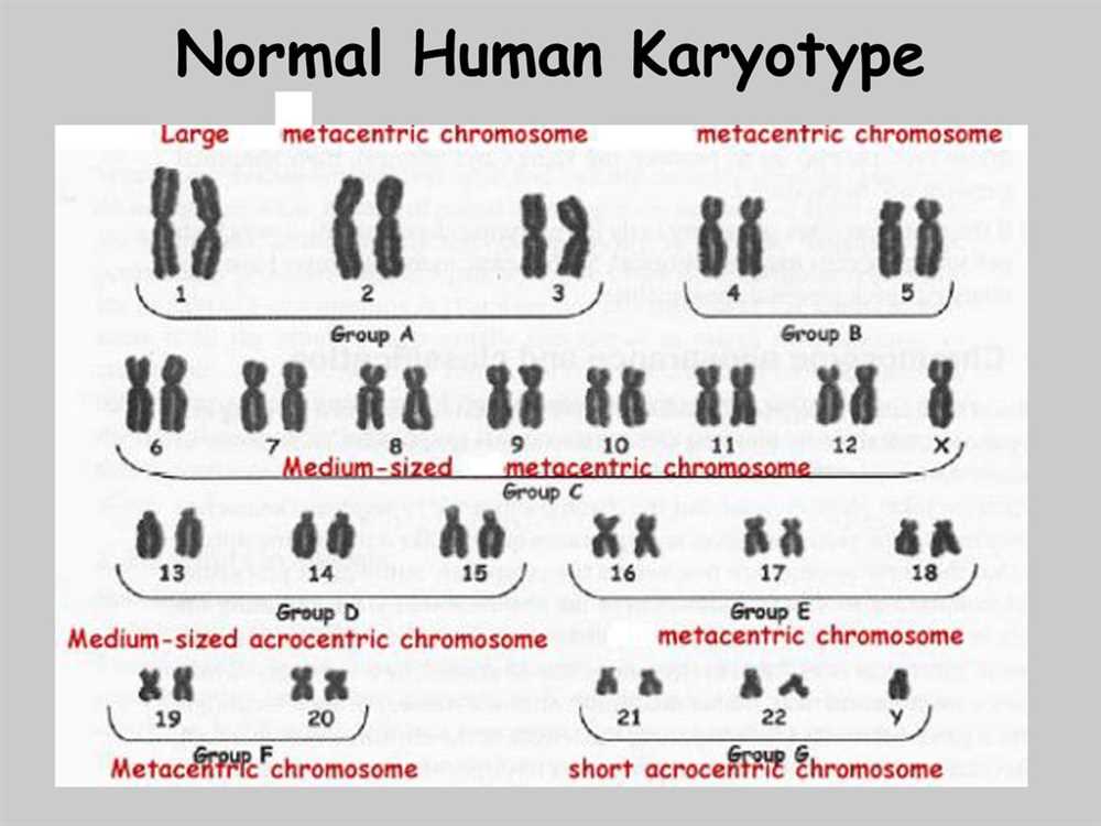
The human karyotype typically consists of 46 chromosomes. These chromosomes are categorized into two types: autosomes and sex chromosomes. Autosomes are the 22 pairs of chromosomes that are the same in both males and females, while sex chromosomes determine the sex of an individual, with females having two X chromosomes (XX) and males having one X and one Y chromosome (XY).
The study and analysis of human karyotypes are essential in various fields, including genetics, medicine, and forensics. They provide valuable information about an individual’s genetic makeup, can help diagnose genetic diseases, and aid in the identification of individuals in criminal investigations or paternity cases.
Importance of understanding human karyotypes
The study of human karyotypes plays a crucial role in understanding various genetic disorders and diseases. By examining the structure and organization of chromosomes within an individual’s cells, scientists can gain valuable insights into the presence of abnormal genetic conditions. Karyotyping not only helps in the diagnosis of genetic disorders but also aids in determining their specific nature and severity.
A deep understanding of human karyotypes allows healthcare professionals to provide accurate genetic counseling and personalized treatment plans. By identifying chromosomal abnormalities, such as the presence of an extra or missing chromosome, doctors can predict potential health issues and help patients make informed decisions regarding family planning and reproductive choices. Moreover, karyotyping assists in determining the risk of passing on genetic disorders to future generations, enabling couples to make informed choices about having children.
Additionally, studying human karyotypes is essential for researchers and scientists in their pursuit of advancing medical knowledge. The identification of specific chromosomal abnormalities associated with certain diseases or conditions can lead to the development of targeted therapies and treatments. Through karyotyping, scientists can also gain a better understanding of the genetic basis of diseases, allowing for the development of more accurate diagnostic tests in the future.
In conclusion, understanding human karyotypes is of paramount importance for both medical professionals and researchers. It not only aids in the diagnosis and management of genetic disorders but also contributes to the advancement of medical knowledge, ultimately leading to improved patient care and treatment outcomes.
Step-by-step guide to the human karyotype exercise
In the human karyotype exercise, students are given a set of chromosomes and must cut them out and arrange them in the correct order to create a karyotype. Karyotyping is the process of organizing chromosomes into pairs and analyzing their structure. This exercise is often used to teach students about genetic disorders and how they can be detected through chromosomal abnormalities.
1. Gather the necessary materials:
- Printed copies of the chromosome set
- Scissors
- Glue or tape
- Blank paper or poster board
2. Familiarize yourself with the chromosomes:

Before you begin cutting out the chromosomes, take some time to study the set and become familiar with the different shapes and sizes of the chromosomes. This will help you identify and arrange them correctly later on.
3. Cut out the chromosomes:
Carefully use the scissors to cut out each individual chromosome from the set. Be sure to cut along the outline of each chromosome, trying to keep as close to the line as possible to maintain their accuracy.
4. Arrange the chromosomes:
On a blank piece of paper or poster board, start arranging the chromosomes in pairs according to their size and banding patterns. Remember that chromosomes are arranged in pairs, with one coming from the mother and one from the father.
5. Glue or tape the chromosomes in place:
Once you have arranged the chromosomes in the correct pairs, use glue or tape to secure them in place on the paper or poster board. Make sure each pair is properly labeled with its corresponding chromosome number.
6. Analyze the karyotype:
Take a step back and examine your completed karyotype. Look for any abnormalities or irregularities in the chromosome pairs. These can indicate genetic disorders such as Down syndrome or Turner syndrome.
By following this step-by-step guide, students can successfully complete the human karyotype exercise and gain a better understanding of the structure and arrangement of chromosomes in the human genome.
Collecting the necessary materials
When performing a human karyotype exercise, it is important to gather all the necessary materials beforehand. Having everything prepared will ensure a smooth and efficient process.
Microscope: The first essential item is a microscope, specifically a compound microscope with high magnification capabilities. A microscope allows for a detailed examination of the chromosomes and enables the identification of any structural abnormalities.
Slide and cover slips: Slides and cover slips are used to hold the specimen in place and protect it during the examination process. It is important to ensure that the slides and cover slips are clean and free from any debris or contaminants.
Stain: Staining the chromosomes is essential to enhance their visibility under the microscope. Various stains, such as Giemsa stain, can be used to highlight the unique banding patterns of the chromosomes, aiding in their identification.
Cell culture: To obtain the necessary cells for karyotyping, a cell culture is required. This involves collecting a sample of cells, such as from amniotic fluid or blood, and culturing them in a nutrient-rich medium to encourage cell division.
Cutting tools: Fine-pointed scissors and forceps are necessary for the physical manipulation of the specimen. These tools are used to carefully cut and isolate individual chromosomes for examination.
Reference charts: Reference charts, such as a karyotype chart or an atlas of human cytogenetics, are invaluable resources during the exercise. They provide a visual representation of normal and abnormal human karyotypes, allowing for comparison and identification.
Notebook and pen: Keeping a notebook and pen handy is essential for documenting observations and recording any abnormalities or significant findings during the exercise. It is important to maintain detailed records for reference and analysis.
By collecting and organizing all the necessary materials, the human karyotype exercise can be conducted effectively, leading to a better understanding of chromosomal abnormalities and their impact on human health.
Sample Preparation
Sample preparation is a crucial step in the process of analyzing human karyotypes. It involves several key procedures that are necessary to ensure successful results. One of the main objectives of sample preparation is to obtain cells from the patient’s body that can be examined under a microscope. These cells are typically collected from sources such as blood, bone marrow, or tissues.
Cell Culturing: In order to obtain a sufficient number of cells for analysis, it is often necessary to culture them in the laboratory. Cell culturing involves providing cells with the necessary nutrients and conditions for growth and multiplication. This can be achieved by placing the collected cells in a culture medium that contains essential nutrients, such as amino acids, vitamins, and growth factors. Controlled temperature and humidity are also important factors to maintain for the growth of healthy cells.
Cell Fixation: After obtaining a sufficient number of cells, the next step is to fix them. Cell fixation involves treating the cells with a fixative solution that preserves their structure and prevents decay. The most commonly used fixative solution is a mixture of methanol and acetic acid, known as the Carnoy’s fixative. This solution helps to cross-link proteins and nucleic acids within the cells, making them more resistant to degradation during subsequent steps.
Chromosome Spreading: Once the cells are fixed, they need to be spread onto a glass slide in order to visualize their chromosomes. This can be achieved by carefully dropping a small amount of the cell suspension onto the slide and allowing it to air-dry. The slide is then ready for further processing and staining.
Staining: Staining is an essential step in sample preparation as it helps to distinguish the different chromosomes and visualize their structural abnormalities. Two common staining techniques used in karyotyping are Giemsa staining and fluorescent in situ hybridization (FISH). Giemsa staining produces a unique pattern of bands on the chromosomes, which helps in identifying specific regions. FISH, on the other hand, uses fluorescently labeled DNA probes to target specific genes or chromosomal regions, allowing for more precise analysis of genetic abnormalities.
Overall, sample preparation plays a critical role in obtaining high-quality images of chromosomes for karyotype analysis. By carefully following these procedures, researchers can ensure accurate and reliable results that can help in the diagnosis of various genetic disorders.
Cut and Paste Method
The cut and paste method is a common technique used in various fields, including biology and genetics, to manipulate genetic materials for analysis and study. This method involves physically cutting out segments of DNA or chromosomes and pasting them onto a desired substrate, such as a microscope slide or a karyotype diagram.
The cut and paste method is particularly useful in studying human karyotypes, which are visual representations of an individual’s chromosomes. Karyotyping involves arranging and pairing the chromosomes to identify any abnormalities or genetic disorders.
To create a human karyotype using the cut and paste method, a biologist would start by collecting cells from a person’s body, typically through a biopsy or blood sample. These cells are then treated to stop them in the metaphase stage of cell division, where the chromosomes are most condensed and visible under a microscope.
The next step involves preparing a karyotype diagram, which consists of blank spaces representing the chromosomes. With the help of specialized tools, the biologist cuts out each chromosome from the microscope slide and carefully pastes them onto the corresponding spaces on the karyotype diagram.
Once all the chromosomes are placed on the diagram, the biologist can analyze and study the karyotype to identify any abnormalities or genetic disorders. This method allows for a clearer visualization and comparison of chromosomes, making it easier to detect any structural or numerical abnormalities.
Analyzing the results
After completing the human karyotype exercise, the results can be analyzed to gain insights into the genetic makeup of an individual. The karyotype is a visual representation of an individual’s chromosomes, which allows scientists to study the number, size, and shape of the chromosomes. By examining the karyotype, we can identify any abnormalities or variations that may be present.
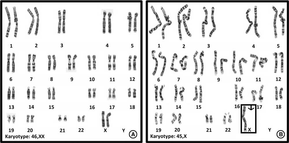
One of the main aspects to consider when analyzing the results is the sex of the individual. In a normal human karyotype, females typically have two X chromosomes, while males have one X and one Y chromosome. Any deviations from this pattern can indicate genetic disorders or conditions, such as Turner syndrome or Klinefelter syndrome.
Another important aspect to look for is the presence of any structural abnormalities in the chromosomes. This can include deletions, duplications, translocations, or inversions. These structural abnormalities can be associated with genetic disorders, such as Down syndrome, Cri-du-chat syndrome, or certain types of leukemia.
Furthermore, the size and shape of the chromosomes can provide additional information about the genetic health of the individual. Variations in chromosomal banding patterns can indicate genetic mutations or rearrangements, which may contribute to the development of certain diseases.
In conclusion, analyzing the results of the human karyotype exercise allows us to identify any abnormalities, variations, or genetic disorders that may be present in an individual. This information is vital for understanding the individual’s genetic health and can aid in diagnosing and treating genetic conditions.