In the field of radiology, the ability to identify and interpret X-rays is a crucial skill. X-rays are a type of electromagnetic radiation used to produce images of the internal structures of the body. They play a vital role in the diagnosis and treatment of various medical conditions.
Haspi 08d is a series of questions that test students’ knowledge and understanding of X-ray interpretation. These questions are designed to challenge students’ abilities to identify and analyze different types of X-rays, such as chest X-rays, head X-rays, and skeletal X-rays.
By answering the Haspi 08d questions, students can gain a deeper understanding of the anatomy and pathology associated with different medical conditions. They can learn to identify the presence of fractures, tumors, or other abnormalities in X-ray images and make informed judgments about the appropriate course of action.
This article provides comprehensive answers to the Haspi 08d questions, guiding students through the process of identifying and interpreting X-rays in a systematic and logical manner. By mastering the skills required to analyze X-rays effectively, students can develop the foundation necessary for a successful career in radiology.
What is x-ray imaging and its importance in medical diagnosis?
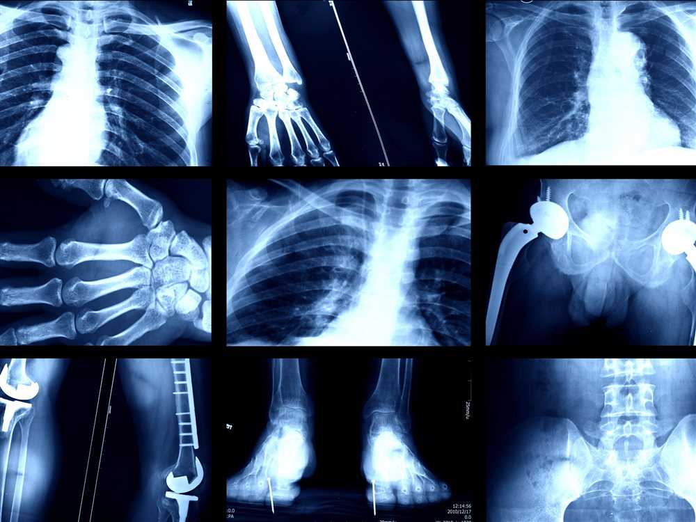
X-ray imaging, also known as radiography, is a commonly used medical imaging technique that allows doctors to visualize the internal structures of the body. It involves the use of a small amount of ionizing radiation to create detailed images of bones, organs, and tissues. X-rays are a type of electromagnetic radiation that can easily pass through soft tissues, but are absorbed by denser materials such as bones, creating contrasting images on a film or digital detector. This non-invasive procedure is commonly used to diagnose and monitor a wide range of medical conditions.
X-ray imaging plays a crucial role in medical diagnosis due to its ability to provide valuable information about the presence of abnormalities or diseases. It allows doctors to detect and diagnose fractures, bone infections, tumors, lung conditions, gastrointestinal issues, and various other conditions. It is particularly effective in identifying skeletal abnormalities, such as fractures or dislocations, as well as identifying the presence of foreign objects in the body. X-ray images can also help guide medical interventions, such as placing catheters or other devices in the correct position.
The importance of x-ray imaging lies in its ability to provide quick and accurate information that can aid in the diagnosis and treatment of patients. X-rays are relatively inexpensive, widely available, and can be performed quickly, making them an efficient tool for medical professionals. They are often the first imaging technique used in emergency situations to assess injuries or trauma. X-rays can help determine the severity of an injury, guide the appropriate treatment plan, and monitor the progress of healing over time. This imaging technique is also useful for screening purposes, allowing doctors to detect diseases at an early stage when treatment options may be more effective.
In conclusion, x-ray imaging is a valuable and widely used medical imaging modality that provides important diagnostic information. It allows doctors to visualize internal structures, detect abnormalities, and guide medical interventions. With its accessibility, efficiency, and ability to provide crucial information, x-ray imaging plays a vital role in medical diagnosis and patient care.
Overview of x-ray imaging technology

X-ray imaging is a widely used medical imaging technique that allows doctors to see inside the body in order to diagnose and treat various medical conditions. It works by using x-ray machines to generate high-energy electromagnetic radiation, which passes through the body and is captured on a receptor, such as a film or a digital detector. This creates an image that can be viewed by medical professionals to identify abnormalities or injuries.
X-ray imaging technology has evolved significantly since its discovery in the late 19th century. Today, digital x-ray systems are commonly used, replacing traditional film-based systems. These digital systems offer numerous advantages, including faster image acquisition, improved image quality, and the ability to manipulate and enhance images for better visualization. Digital x-ray images can be easily stored and shared electronically, allowing for quick and convenient access by healthcare professionals.
The use of x-ray imaging is not limited to just medical applications. It is also widely utilized in non-destructive testing and security screening. In non-destructive testing, x-ray imaging is used to inspect the structural integrity of materials, such as metals or composites, without causing damage. In security screening, x-ray scanners are used to detect suspicious objects or substances hidden within luggage or containers.
While x-ray imaging is a valuable tool in healthcare and other industries, it is important to note that it does involve exposure to ionizing radiation. Therefore, appropriate safety measures must be taken to minimize radiation dose to the patient and healthcare personnel. This includes the use of lead shielding and collimation techniques to limit unnecessary radiation exposure.
Advantages of x-ray imaging technology:
- Allows for non-invasive visualization of internal structures
- Quick and convenient imaging process
- Digital systems offer improved image quality and manipulation capabilities
- Can be used in various applications, such as medical diagnosis, non-destructive testing, and security screening
Disadvantages of x-ray imaging technology:

- Involves exposure to ionizing radiation
- May have limitations in visualizing certain tissues or structures
- Can be costly to implement and maintain
- Requires trained professionals to operate the equipment and interpret the images
Role of x-ray imaging in diagnosing medical conditions
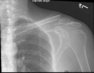
X-ray imaging plays a crucial role in diagnosing various medical conditions and it is an essential tool used by healthcare professionals. X-rays are a form of electromagnetic radiation that can penetrate the body and create images of the internal structures. They are commonly used to diagnose bone fractures, dislocations, and joint abnormalities. X-ray images provide valuable information about the location and severity of injuries, helping doctors plan appropriate treatment strategies.
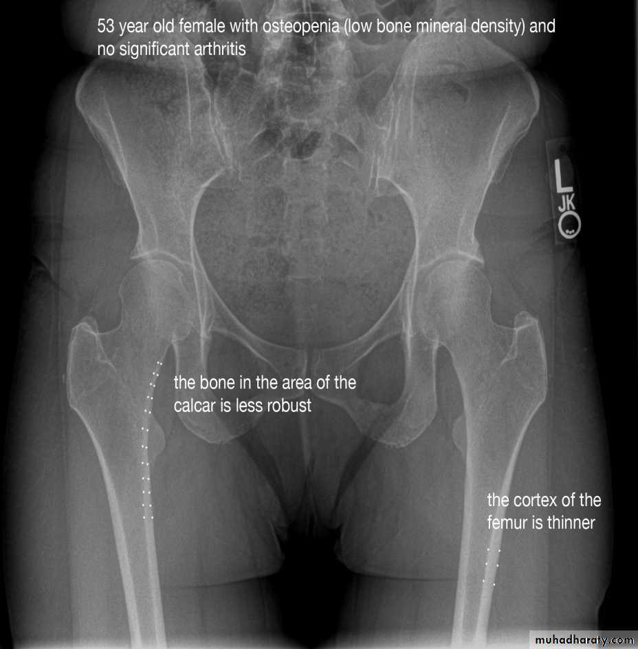
One of the key advantages of x-ray imaging is its ability to detect abnormalities in the skeletal system. It is particularly useful in identifying fractures, which are often difficult to diagnose through physical examination alone. X-ray images of the affected area can reveal the extent of the injury, the alignment of the bones, and the presence of any associated complications such as bone fragments or dislocations. Furthermore, x-rays can also diagnose conditions like osteoporosis, bone tumors, and degenerative joint diseases, providing valuable insights for appropriate treatment.
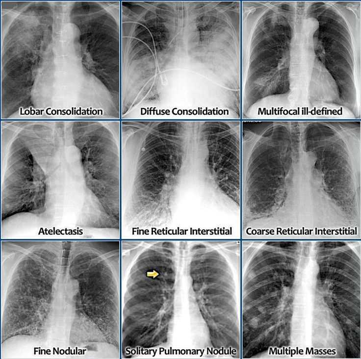
X-rays are also commonly used to diagnose conditions affecting the respiratory system. They are an important tool in detecting lung diseases such as pneumonia, tuberculosis, and lung cancer. X-ray imaging can reveal abnormalities in the lung tissue, such as inflammation, fluid accumulation, or the presence of tumors. It can also help evaluate the size and position of the heart and detect conditions like congestive heart failure or enlarged blood vessels in the lungs.
Moreover, x-ray imaging is frequently used in the diagnosis of gastrointestinal conditions. It is an efficient way to visualize the digestive organs such as the stomach, intestines, and colon, allowing doctors to identify obstructions, perforations, or abnormalities in the gastrointestinal tract. X-rays can also help diagnose conditions like kidney stones or urinary tract infections, by identifying any calcifications or abnormalities in the urinary system.
In conclusion, x-ray imaging is a vital diagnostic tool that plays a significant role in identifying and diagnosing various medical conditions. Its ability to provide detailed images of the skeletal system, respiratory system, and gastrointestinal system allows healthcare professionals to accurately diagnose and treat patients, ensuring appropriate care and management of their conditions.
Understanding the characteristics of x-ray images
X-ray images play a vital role in the diagnostic process of various medical conditions. These images are created by passing x-ray beams through the body, and they provide valuable information about the internal structures and organs. To interpret x-ray images accurately, it is essential to understand their characteristics and key features.
- Density variations: X-ray images display various levels of density, ranging from air to bone. Dense structures, such as bones, appear white in x-ray images due to their high density, while less dense structures, such as air-filled cavities, appear black. Soft tissues, which have intermediate density, appear as shades of gray. This density variation allows healthcare professionals to differentiate between different anatomical structures.
- Anatomical details: X-ray images provide detailed information about the anatomical structures, such as bones, organs, and blood vessels. By analyzing the shape, size, and position of these structures, medical professionals can identify abnormalities, fractures, tumors, and other conditions that may affect the patient’s health. This enables accurate diagnosis and appropriate treatment planning.
- Contrast and exposure: Proper contrast and exposure settings are crucial for obtaining high-quality x-ray images. Contrast refers to the difference in brightness between the different shades of gray, and exposure determines the overall darkness or brightness of the image. Adequate contrast and exposure settings ensure clear visualization of the anatomical structures and help identify any abnormalities present.
- Artifacts: X-ray images may sometimes contain artifacts, which are unintended structures or distortions that can affect the interpretation of the image. Common artifacts include motion blur, scatter radiation, and image noise. It is important to identify and minimize these artifacts to obtain accurate diagnostic information from the x-ray images.
Overall, understanding the characteristics of x-ray images is essential for healthcare professionals to interpret the images correctly and make accurate diagnoses. Through proper analysis of density variations, anatomical details, contrast and exposure settings, and identification of artifacts, medical professionals can leverage the valuable information provided by x-ray images to deliver effective patient care.
How do x-rays work?

X-rays are a type of electromagnetic radiation with a short wavelength and high energy. They were discovered in 1895 by Wilhelm Conrad Roentgen, who observed that they could pass through materials that were opaque to visible light. Since then, x-rays have become an essential tool in various fields, including medicine, industry, and research.
When x-rays pass through the human body, they can be absorbed, scattered, or pass straight through depending on the type of tissues they encounter. Dense tissues, such as bones, absorb more x-rays and appear white on the resulting image, whereas less dense tissues, like muscles or organs, absorb fewer x-rays and appear darker. This differential absorption allows medical professionals to identify abnormalities or fractures in bones, detect tumors, and diagnose various health conditions.
Inside an x-ray machine, a controlled beam of x-rays is generated by an x-ray tube. The tube contains a cathode and an anode, between which a high voltage is applied. When the cathode is heated, it emits electrons, which are accelerated towards the anode. As the electrons collide with the anode, x-ray photons are produced. The x-ray photons then pass through the patient’s body and are detected by an image receptor on the other side. The information collected by the image receptor is then processed to create an x-ray image that can be analyzed by medical professionals.
Although x-rays are highly useful in medical imaging, they do carry some risks. Exposure to high levels of x-ray radiation can damage cells and DNA, potentially leading to cancer or other health issues. Therefore, precautions are taken to limit the amount of radiation patients and medical personnel are exposed to during x-ray procedures. Lead aprons and collars are often worn to shield the body from unnecessary exposure, and x-ray machines are carefully calibrated to deliver the minimum amount of radiation necessary to obtain the required diagnostic information.
In conclusion, x-rays work by emitting a controlled beam of high-energy electromagnetic radiation, which passes through the body and generates an image by differential absorption. They are a valuable tool in medicine and other fields but should be used with caution to avoid potential health risks associated with radiation exposure.
Key characteristics of x-ray images
X-ray images, also known as radiographs, provide valuable diagnostic information in the field of medical imaging. These images are produced by passing X-ray radiation through the body and capturing the resulting energy with a specialized detector. X-ray images have several key characteristics that make them an essential tool in medical diagnosis and treatment.
1. Density variations
One of the main features of x-ray images is the ability to visualize density variations within the body. Different tissues and organs have varying densities, which can be seen as differences in shades of gray on the image. For example, bones appear white because they absorb a high amount of X-rays, while air-filled structures like the lungs appear black because they allow most of the X-rays to pass through. This contrast in density enables clinicians to identify abnormalities such as fractures, tumors, and fluid accumulation.
2. Structural details
X-ray images provide detailed information about the internal structures of the body. This allows healthcare professionals to assess the condition of bones, joints, and internal organs. For instance, fractures can be detected by analyzing the alignment and continuity of the bone structure. X-ray images can also reveal structural abnormalities, such as the enlargement of organs or the presence of foreign objects, helping in the diagnosis of various conditions.
3. Real-time imaging
X-ray imaging is a real-time technique, meaning that the images can be obtained instantaneously. This makes it particularly useful in emergency situations, where immediate diagnosis and treatment are necessary. X-rays can capture dynamic processes such as the movement of bones or the flow of contrast agents through blood vessels. Real-time x-ray imaging, also known as fluoroscopy, is often used during procedures such as cardiac catheterization or joint injections.
4. Low cost and accessibility

X-ray imaging is relatively inexpensive compared to other diagnostic imaging modalities such as MRI or CT scans. X-ray machines are widely available in healthcare facilities, making the technology accessible to a large population. This affordability and accessibility contribute to the widespread use of x-ray imaging in various medical settings, from small clinics to large hospitals.
In conclusion, x-ray images have key characteristics that make them an indispensable tool in medical diagnosis. The ability to visualize density variations, assess structural details, provide real-time imaging, and the low cost and accessibility make x-ray imaging an important component of modern healthcare.