
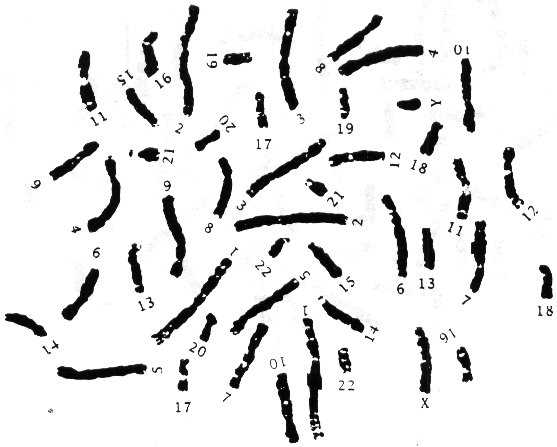
In the field of genetics, studying karyotypes is essential in understanding chromosomal disorders. A karyotype is a visual representation of an individual’s chromosomes, organized according to their size, shape, and banding patterns. By analyzing karyotypes, scientists can identify structural abnormalities and numerical disorders that may lead to genetic disorders.
One way to understand karyotypes is through a webquest, where students are tasked with analyzing different karyotypes and answering questions based on their findings. This answer key will provide a comprehensive guide on how to interpret karyotypes and understand the various chromosomal disorders that may be present.
With this answer key, students can learn about common chromosomal disorders such as Down syndrome, Turner syndrome, and Klinefelter syndrome. Additionally, they will gain a deeper understanding of how chromosomal abnormalities can impact an individual’s physical and cognitive development.
Karyotype Webquest Answer Key
In the field of genetics, karyotyping is a valuable tool used to analyze an individual’s chromosomes. A karyotype is a visual representation of the chromosomes arranged in pairs and organized according to their size, shape, and banding patterns. By examining a karyotype, scientists can identify any abnormalities or changes in the number or structure of chromosomes.
During the Karyotype Webquest, students were tasked with analyzing different karyotypes and answering specific questions related to each one. Here is the answer key that provides the correct responses:
Question 1:
What is the sex of the individual represented in Karyotype A?
The individual represented in Karyotype A is a male (XY). The presence of a Y chromosome indicates the male sex.
Question 2:
How many chromosomes are found in Karyotype B?
Karyotype B contains a total of 46 chromosomes. This is the normal number of chromosomes found in a human karyotype.
Question 3:
Identify any abnormalities or changes in the karyotype displayed in Karyotype C.
The karyotype displayed in Karyotype C shows an extra copy of chromosome 21, resulting in a condition known as trisomy 21 or Down syndrome. This extra chromosome leads to the characteristic physical and intellectual disabilities seen in individuals with Down syndrome.
Question 4:
What is the sex of the individual represented in Karyotype D?
The individual represented in Karyotype D is a female (XX). The presence of two X chromosomes indicates the female sex.
By completing the Karyotype Webquest and using the answer key provided, students can enhance their understanding of karyotyping and its importance in genetic analysis. The webquest allows them to practice interpreting karyotypes and identifying any abnormalities or changes that may be present. This activity helps deepen their knowledge of genetics and prepares them for future studies in the field.
What is a Karyotype?
A karyotype is a visual representation of an individual’s chromosomes. Chromosomes are the structures within cells that carry genetic information, and they can be observed under a microscope. A karyotype provides a way to examine an individual’s chromosomes and identify any abnormalities or variations.
To create a karyotype, cells from the individual are first collected and grown in a lab. Then, the cells are stopped during cell division, usually in metaphase, when the chromosomes are most condensed and visible. The chromosomes are stained and arranged in pairs, according to their size, shape, and banding patterns.
The karyotype provides valuable information about an individual’s genetic makeup. It can reveal the number and structure of chromosomes, as well as identify any chromosomal abnormalities, such as extra or missing chromosomes, translocations, deletions, or duplications. This information is important for diagnosing genetic disorders, determining the sex of an individual, and assessing the risk of certain genetic conditions.
The karyotype is typically presented as a chart, with pairs of chromosomes arranged in numerical order. The first 22 pairs are called autosomes, and the 23rd pair is the sex chromosomes, which determine the individual’s biological sex. In a normal karyotype, females have two X chromosomes (XX), while males have one X and one Y chromosome (XY).
Overall, a karyotype is a powerful tool in genetics and cytogenetics that allows scientists and healthcare professionals to examine an individual’s chromosomes and understand their genetic profile.
Importance of Karyotyping in Genetics
Karyotyping is an essential tool in the field of genetics as it allows scientists to visualize and analyze an individual’s chromosomes. By examining the structure, number, and arrangement of chromosomes, karyotyping provides valuable information about an individual’s genetic makeup and can identify any abnormalities or genetic disorders.
Karyotyping plays a crucial role in identifying and diagnosing chromosomal abnormalities such as Down syndrome, Turner syndrome, and Klinefelter syndrome. These abnormalities can have significant impacts on a person’s physical, intellectual, and developmental abilities. By performing a karyotype analysis, healthcare professionals can accurately diagnose these conditions and provide appropriate medical interventions and support.
Karyotyping is also essential in the field of reproductive genetics. It helps identify chromosomal abnormalities in embryos during prenatal testing, allowing parents to make informed decisions about their pregnancy. Additionally, karyotyping is used in pre-implantation genetic diagnosis (PGD) during in vitro fertilization (IVF) procedures to select embryos without chromosomal abnormalities for implantation, increasing the chances of a successful pregnancy.
The study of karyotypes in cancer is another important application of karyotyping in genetics. Cancer cells often exhibit abnormal karyotypes, characterized by chromosomal rearrangements, duplications, deletions, or translocations. These changes can lead to the development and progression of cancer. By analyzing cancer karyotypes, researchers can identify the specific chromosomal alterations driving cancer growth, which can inform the development of targeted therapies and personalized treatment approaches.
In summary, karyotyping is an invaluable tool in genetics that allows for the identification of chromosomal abnormalities, the diagnosis of genetic disorders, and the study of cancer karyotypes. Its applications extend across various areas of genetics and have significant implications for patient care, reproductive medicine, and cancer research.
Steps in the Karyotyping Process
The process of karyotyping involves several steps in order to analyze the chromosomes and identify any abnormalities or genetic disorders. These steps are essential in understanding the genetic composition of an individual and can provide valuable information for diagnosis and treatment.
1. Sample Collection: The first step in the karyotyping process is to collect a sample containing the individual’s cells, such as blood or amniotic fluid. The sample is usually collected using a non-invasive method and then transported to the laboratory for further analysis.
2. Cell Culturing: Once the sample is collected, the cells need to be cultured in a laboratory setting. This involves providing the cells with nutrients and ideal conditions for growth and multiplication. By culturing the cells, there will be enough material to examine and analyze the chromosomes.
3. Chromosome Staining: After cell culturing, the next step is to stain the chromosomes. Staining techniques allow the chromosomes to be visible under a microscope and enable their identification and analysis. The most commonly used staining method is called Giemsa staining, which produces characteristic bands on the chromosomes.
4. Chromosome Analysis: Once the chromosomes are stained, they can be examined and analyzed. They are arranged based on their size, structure, and banding pattern. This process is called karyotyping, and it allows for the identification of any alterations or abnormalities in the chromosomes, such as deletions, duplications, translocations, or changes in the number of chromosomes.
5. Result Interpretation: The final step in the karyotyping process is interpreting the results. A trained geneticist or cytogeneticist examines the karyotype and determines if there are any abnormal findings. They compare the individual’s karyotype to a normal reference karyotype and look for any discrepancies. This interpretation is crucial for accurate diagnosis and providing appropriate medical recommendations.
In conclusion, the karyotyping process involves sample collection, cell culturing, chromosome staining, chromosome analysis, and result interpretation. These steps are essential in understanding the structure and organization of an individual’s chromosomes and identifying any genetic abnormalities or disorders. By following these steps, healthcare professionals can provide accurate diagnoses and develop appropriate treatment plans for patients.
Types of Chromosomal Abnormalities
Chromosomal abnormalities can occur in different forms, leading to a range of genetic disorders and conditions. These abnormalities can affect the structure, number, or arrangement of chromosomes in an individual’s cells. Here are some common types of chromosomal abnormalities:
Nondisjunction
Nondisjunction is a type of chromosomal abnormality where chromosomes fail to separate properly during cell division. This can result in the formation of cells with an abnormal number of chromosomes. For example, individuals with Down syndrome have an extra copy of chromosome 21 due to nondisjunction. Nondisjunction can occur during meiosis, leading to an abnormal number of chromosomes in the gametes, which can then result in an abnormal number of chromosomes in the offspring.
Deletion
In a deletion chromosomal abnormality, a portion of a chromosome is missing. This can occur due to a breakage in a chromosome, and when the broken part is lost during repair, it results in a deletion. The loss of genetic material can lead to various health issues, depending on the specific genes affected by the deletion. For example, individuals with Williams syndrome have a deletion on chromosome 7, leading to developmental delays and characteristic facial features.
Duplication
Duplication occurs when a section of a chromosome is duplicated, resulting in an extra copy of genetic material. This can happen through errors during DNA replication or other genetic processes. Duplication can have varying effects depending on the genes involved. In some cases, it may lead to developmental delays or other health issues.
Inversion
In an inversion chromosomal abnormality, a segment of a chromosome is flipped in orientation. This means that the genes within the inverted segment are in the opposite order compared to the normal arrangement. Inversions can occur without any apparent health consequences, but they can also disrupt the normal functioning of genes and lead to genetic disorders.
Translocation
Translocation involves the exchange of genetic material between two non-homologous chromosomes. This can result in rearrangements of genetic material and can be balanced or unbalanced. In balanced translocations, the total amount of genetic material remains unchanged, but the arrangement is altered. Unbalanced translocations, on the other hand, can lead to an abnormal amount of genetic material and can cause various developmental issues.
These are just a few examples of the different types of chromosomal abnormalities that can occur. Each type of abnormality can have specific effects on an individual’s health and development, and understanding them is important for diagnosing and managing genetic disorders and conditions.
Interpreting a Karyotype
Interpreting a karyotype is a crucial step in understanding an individual’s genetic makeup. A karyotype is a visual representation of a person’s chromosomes, which are organized and arranged according to their size, shape, and banding pattern. Each chromosome is paired with its corresponding homologous chromosome, resulting in a total of 23 pairs of chromosomes in a human karyotype.
To interpret a karyotype, one must first observe the overall structure and organization of the chromosomes. Normal karyotypes typically show two sets of chromosomes, with each pair labeled from 1 to 22, known as autosomes, and the 23rd pair representing the sex chromosomes, labeled as either XX for female or XY for male. Abnormalities in the number or structure of chromosomes can indicate genetic disorders or conditions.
By examining the karyotype, it is possible to identify various chromosome abnormalities, such as trisomy, monosomy, or translocations. Trisomy refers to the presence of an extra chromosome, while monosomy refers to the absence of a chromosome. Both trisomy and monosomy can have significant implications for an individual’s health and development. Translocations, on the other hand, involve the rearrangement of genetic material between chromosomes.
Common abnormalities observed in a karyotype include:
- Down syndrome (trisomy 21) – an extra copy of chromosome 21 is present.
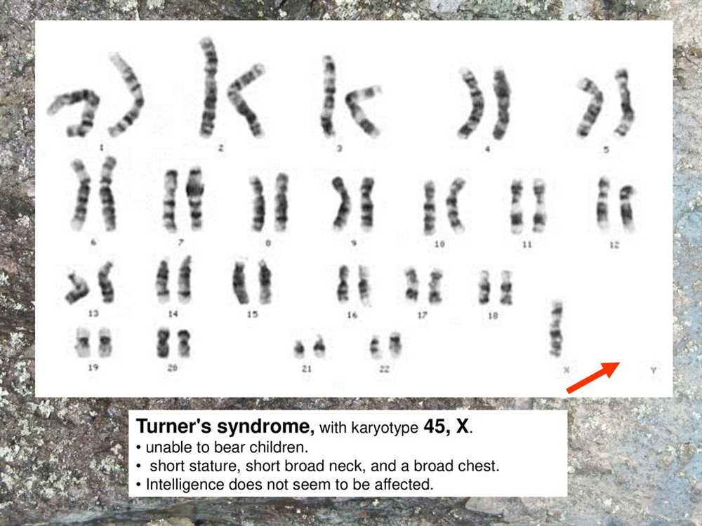
- Turner syndrome (monosomy X) – females have only one X chromosome instead of two.
- Klinefelter syndrome (XXY) – males have an extra X chromosome, resulting in XXY instead of XY.
- Philadelphia chromosome (translocation) – a specific translocation involving chromosomes 9 and 22, often associated with chronic myeloid leukemia.
Overall, interpreting a karyotype provides valuable insights into an individual’s genetic composition and can help diagnose genetic disorders and conditions. It plays a crucial role in genetic counseling, prenatal testing, and medical research.
Common Findings in Karyotypes
Karyotyping is a technique used to analyze the chromosomes of an individual. It allows scientists to identify any abnormalities or variations in the structure or number of chromosomes. While each individual’s karyotype is unique, there are some common findings that can be observed.
1. Normal Karyotype
A normal karyotype consists of 46 chromosomes, arranged in 23 pairs. These pairs are numbered from 1 to 22, with the 23rd pair being the sex chromosomes (XX for females and XY for males). A normal karyotype shows no major structural abnormalities or additional chromosomes.
2. Trisomies
Trisomies occur when there is an extra copy of a particular chromosome. The most well-known trisomy is Trisomy 21, also known as Down syndrome. In this condition, there is an extra copy of chromosome 21, resulting in characteristic physical features and intellectual disabilities. Other trisomies, such as Trisomy 18 (Edward syndrome) and Trisomy 13 (Patau syndrome), can also be identified through karyotyping.
3. Structural Abnormalities
Structural abnormalities in chromosomes can be observed in karyotypes. These include deletions, duplications, inversions, and translocations. Deletions occur when a portion of a chromosome is missing, while duplications involve an extra copy of a portion of a chromosome. Inversions refer to a segment of a chromosome being flipped in orientation, and translocations involve the rearrangement of genetic material between different chromosomes. These structural abnormalities can have various effects, ranging from mild to severe, depending on the location and size of the alteration.
4. Sex Chromosome Abnormalities
Sex chromosome abnormalities can also be detected through karyotyping. Some common examples include Turner syndrome, in which females only have one X chromosome (45,X) instead of the usual two (46,XX), and Klinefelter syndrome, in which males have an additional X chromosome (47,XXY). Other variations in sex chromosome number, such as XYY and XXX, can also be identified.
In conclusion, karyotyping is a valuable tool in identifying and diagnosing chromosomal abnormalities. By examining the karyotype, scientists can detect common findings such as normal karyotypes, trisomies, structural abnormalities, and sex chromosome abnormalities. Understanding these findings can provide important insights into the genetic makeup and potential health conditions of an individual.