
Skeletal muscles are a complex form of tissue found in the human body, responsible for voluntary movements. Understanding the microscopic anatomy of skeletal muscles is crucial in comprehending their function and how they enable movement.
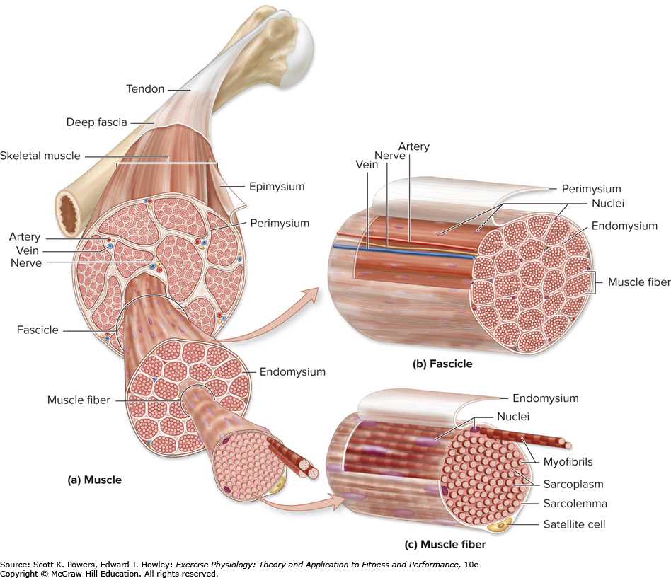
The microscopic anatomy of skeletal muscles includes several key components. One of these components is the muscle fiber, also known as a muscle cell. Muscle fibers are long, cylindrical cells that contain multiple nuclei and are enclosed within a plasma membrane called the sarcolemma.
Within the muscle fiber, there are myofibrils, which are responsible for muscle contractions. Myofibrils are made up of repeating units called sarcomeres. Sarcomeres consist of two main protein filaments, actin and myosin, which are responsible for the sliding filament theory of muscle contraction.
In addition to myofibrils, skeletal muscles also contain specialized structures called T-tubules and sarcoplasmic reticulum. T-tubules are invaginations of the sarcolemma that allow for the transmission of electrical signals deep into the muscle fiber. The sarcoplasmic reticulum is a specialized form of smooth endoplasmic reticulum that stores and releases calcium ions, which play a crucial role in muscle contraction.
Microscopic Anatomy of Skeletal Muscle Worksheet Chapter 6 Answers
In Chapter 6 of the microscopic anatomy of skeletal muscle worksheet, we explore the intricate details of skeletal muscle at a microscopic level. This worksheet aims to enhance our understanding of the structure and function of skeletal muscle fibers.
One of the key aspects covered in this chapter is the structure of a skeletal muscle fiber. Skeletal muscle fibers are long, cylindrical cells that contain many specialized organelles and structures. They are composed of myofibrils, which are made up of smaller contractile units called sarcomeres. Each sarcomere consists of thick and thin filaments, which slide past each other during muscle contraction.
A critical topic explored in this worksheet is the process of muscle contraction. Muscle contraction is initiated by the release of calcium ions, which bind to proteins on the thin filaments and expose binding sites for the thick filaments. This allows myosin heads to attach to the actin filaments and pull them towards the center of the sarcomere. As a result, the sarcomeres shorten, leading to muscle contraction.
The microscopic anatomy of skeletal muscle also covers the role of motor neurons in muscle contraction. Motor neurons are specialized nerve cells that innervate skeletal muscle fibers. They transmit electrical signals called action potentials, which stimulate the release of calcium ions and initiate muscle contraction. This worksheet delves into the communication between motor neurons and muscle fibers, highlighting the importance of neuromuscular junctions in this process.
Overall, the microscopic anatomy of skeletal muscle worksheet provides in-depth information about the structure and function of skeletal muscle at a microscopic level. It helps students grasp the intricate details of muscle fibers and their role in muscle contraction. By understanding the microscopic anatomy of skeletal muscle, we can gain a deeper appreciation for the complexity and efficiency of the human musculoskeletal system.
Key Terms and Definitions
Skeletal muscle: Also known as striated muscle, skeletal muscle is a type of muscle tissue that is attached to bones and enables voluntary movements.
Fascicle: A fascicle is a bundle of muscle fibers within a skeletal muscle. The muscle fibers within a fascicle are surrounded by connective tissue called perimysium.
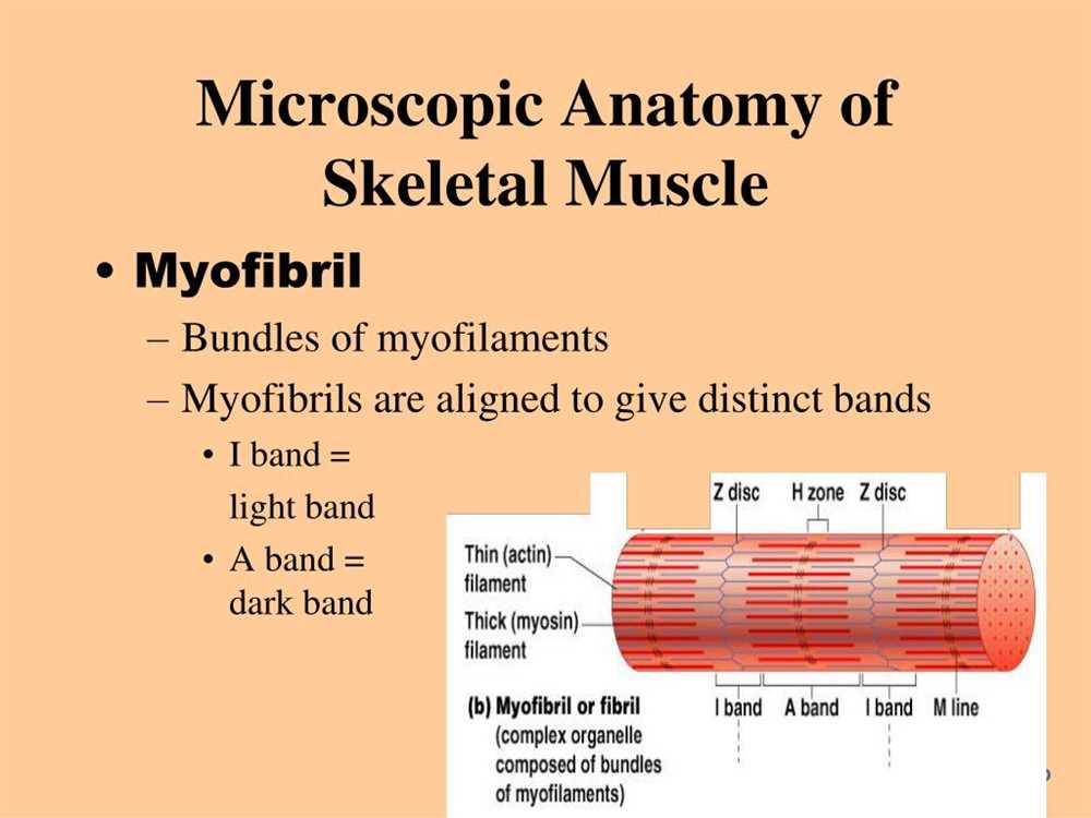
Myofibril: A myofibril is a long, cylindrical structure found within muscle fibers. It is composed of repeating units called sarcomeres, which are responsible for muscle contraction.
Sarcomere: Sarcomeres are the functional units of muscle contraction. They are made up of thin filaments (actin) and thick filaments (myosin) that slide past each other during muscle contraction.
Tendon: A tendon is a tough, fibrous connective tissue that connects muscle to bone. It transmits the force generated by the muscle to produce movement at the joint.
Motor unit: A motor unit consists of a motor neuron and the muscle fibers it innervates. When the motor neuron fires an action potential, all the muscle fibers in the motor unit contract simultaneously.
Neuromuscular junction: The neuromuscular junction is the synapse between a motor neuron and a muscle fiber. It is where the motor neuron releases neurotransmitters, such as acetylcholine, which stimulate muscle contraction.
Endomysium: The endomysium is a thin layer of connective tissue that surrounds individual muscle fibers within a fascicle. It provides support and protection to the muscle fibers.
Sarcoplasmic reticulum: The sarcoplasmic reticulum is a specialized endoplasmic reticulum found in muscle cells. It stores and releases calcium ions, which are necessary for muscle contraction.
T-tubules: T-tubules, also known as transverse tubules, are invaginations of the sarcolemma (muscle cell membrane) that penetrate into the interior of the muscle fiber. They allow for the rapid transmission of electrical impulses from the surface of the muscle fiber to the interior.
Actin: Actin is a globular protein that is a major component of thin filaments in muscle cells. It plays a crucial role in muscle contraction by interacting with myosin to generate force.
Myosin: Myosin is a motor protein that forms the thick filaments in muscle cells. It hydrolyzes ATP to generate the force required for muscle contraction.
Z-line: Z-lines are narrow bands of protein that separate sarcomeres from each other. They anchor the thin filaments and provide structural stability to the sarcomere during muscle contraction.
Structure of Skeletal Muscle Fibers
Skeletal muscles are made up of individual muscle fibers, which are long, cylindrical cells. These fibers contain multiple nuclei and are surrounded by a plasma membrane called the sarcolemma. The sarcolemma plays a vital role in maintaining the structure and function of the muscle fiber, as it regulates the movement of ions in and out of the cell.
Each muscle fiber is composed of smaller units called myofibrils. Myofibrils run parallel to each other within the muscle fiber and are responsible for the contractile properties of the muscle. They are made up of repeating units called sarcomeres, which are the functional units of muscle contraction.
Within each sarcomere, there are two main types of protein filaments: thick filaments made of myosin and thin filaments made of actin, tropomyosin, and troponin. These filaments are arranged in a specific pattern, with the thick filaments located in the center of the sarcomere and the thin filaments extending from either end.
When a muscle contracts, the myosin heads on the thick filaments bind to the actin filaments, causing them to slide past each other. This sliding motion shortens the sarcomere and ultimately leads to muscle contraction.
In addition to the myosin and actin filaments, there are also other proteins involved in the regulation and stabilization of muscle contraction. These include titin, nebulin, and dystrophin, among others. These proteins help to ensure the proper alignment and structure of the muscle fibers during contraction and relaxation.
Overall, the structure of skeletal muscle fibers is highly organized and specialized for contraction. The arrangement of myofibrils, sarcomeres, and protein filaments allows for precise control and coordination of muscle movements.
Sarcomere Structure and Function
The sarcomere is the basic unit of contraction in skeletal muscle. It is a highly organized structure that consists of overlapping thick and thin filaments. The thick filament is composed of myosin protein, while the thin filament is composed of actin protein. The arrangement of these filaments creates a unique banded pattern that can be observed under a microscope.
Within the sarcomere, the A band represents the region of overlap between the thick and thin filaments, while the I band represents the region of only thin filaments. The H zone is the region of only thick filaments. These bands and zones are important for understanding the sliding filament theory of muscle contraction.
During muscle contraction, the sarcomeres shorten as the thick and thin filaments slide past each other. This process is mediated by the interaction between myosin and actin, which forms cross-bridges. The cross-bridges undergo a series of conformational changes, allowing them to pull the thin filaments towards the center of the sarcomere.
Overall, the structure of the sarcomere is vital for muscle contraction. Without the precise arrangement of the thick and thin filaments, the sliding filament mechanism would not be possible. Understanding the sarcomere structure and function is essential for studying the microscopic anatomy of skeletal muscle and its role in movement and other physiological processes.
Myofibril Organization in Skeletal Muscle
Skeletal muscles are made up of bundles of muscle fibers called myofibrils. These myofibrils are responsible for the contraction and movement of skeletal muscles. They are highly organized structures composed of repeating units called sarcomeres.
Each sarcomere is made up of thick myosin filaments and thin actin filaments. The myosin filaments are located in the center of the sarcomere, while the actin filaments are attached to the Z-discs at the ends of the sarcomere. The arrangement of these filaments gives skeletal muscles their striated appearance.
The myosin filaments have protrusions called cross-bridges that attach to the actin filaments. During muscle contraction, these cross-bridges pull on the actin filaments, causing them to slide towards the center of the sarcomere. This shortens the sarcomere and leads to the overall shortening of the muscle fiber.
The organization of myofibrils within skeletal muscles allows for precise control of muscle movements. Each muscle fiber contains multiple myofibrils, and each myofibril contains multiple sarcomeres. When motor neurons stimulate muscle fibers, these sarcomeres contract in a coordinated manner, resulting in the overall contraction of the muscle.
In summary, the organization of myofibrils in skeletal muscles plays a crucial role in muscle contraction and movement. The arrangement of sarcomeres, thick myosin filaments, and thin actin filaments allows for the precise control of muscle contractions, leading to coordinated movements in the body.
Sliding Filament Theory of Muscle Contraction
The sliding filament theory is a widely accepted explanation for how muscles contract. It proposes that muscle contraction occurs when the thin filaments, composed of actin protein, slide past the thick filaments, composed of myosin protein, within muscle cells called sarcomeres. This sliding action shortens the sarcomeres, causing muscle fibers to contract and generate force.
During muscle contraction, the myosin heads on the thick filaments bind to specific sites on the actin molecules of the thin filaments. This binding triggers a conformational change in the myosin heads, causing them to pull the actin filaments towards the center of the sarcomere. This process is facilitated by the hydrolysis of ATP, which provides energy for the myosin heads to move and detach from the actin filaments.
The sliding filament theory also explains how muscle relaxation occurs. When the nervous system signals the muscle to relax, the supply of ATP is depleted and calcium ions are actively transported out of the sarcomere. Without ATP and calcium ions, the myosin heads cannot bind to actin, and the sliding action ceases. The sarcomeres lengthen back to their resting state, and the muscle fibers relax.
Overall, the sliding filament theory provides a detailed understanding of the molecular events that occur during muscle contraction and relaxation. It explains how the interaction between actin and myosin filaments generates force and allows muscles to produce movement. This theory has greatly contributed to our knowledge of skeletal muscle physiology and has paved the way for further research in the field.
Neuromuscular Junction and Muscle Contraction
The neuromuscular junction is a specialized connection between a motor neuron and a skeletal muscle fiber. It is a crucial site for controlling muscle contraction. When a motor neuron reaches the neuromuscular junction, it releases a neurotransmitter called acetylcholine (ACh) into the synaptic cleft. ACh then binds to receptors on the muscle fiber membrane, causing depolarization.
Depolarization of the muscle fiber leads to the production of an action potential, which travels along the sarcolemma and into the T-tubules. This action potential triggers the release of calcium ions from the sarcoplasmic reticulum. The calcium ions bind to troponin, causing a conformational change that moves tropomyosin away from the actin-binding sites.
With the actin-binding sites exposed, myosin heads can bind to actin, forming cross-bridges. The myosin heads then undergo a power stroke, pulling the actin filaments towards the center of the sarcomere. This shortens the sarcomere, resulting in muscle contraction. The power stroke is energized by the hydrolysis of ATP to ADP and inorganic phosphate.
As long as calcium ions and ATP are present, the myosin heads continue to cycle, pulling the actin filaments and generating force. When the motor neuron stops releasing ACh, it is quickly broken down by the enzyme acetylcholinesterase, terminating the depolarization signal. The calcium ions are actively pumped back into the sarcoplasmic reticulum, causing the troponin-tropomyosin complex to return to its blocking position and preventing further muscle contraction.
- The neuromuscular junction is a connection between a motor neuron and a muscle fiber.
- Acetylcholine is the neurotransmitter released by the motor neuron.
- Depolarization of the muscle fiber leads to the production of an action potential.
- The action potential triggers the release of calcium ions from the sarcoplasmic reticulum.
- Calcium ions bind to troponin, moving tropomyosin away from actin-binding sites.
- Myosin heads bind to actin, undergo a power stroke, and pull actin filaments towards the center of the sarcomere.
- This shortens the sarcomere and causes muscle contraction.
- Myosin heads continue to cycle as long as ATP and calcium ions are present.
- When ACh is no longer released, the depolarization signal is terminated.
- Calcium ions are actively pumped back into the sarcoplasmic reticulum, returning the troponin-tropomyosin complex to its blocking position.