
Muscle contraction is a complex process that allows our bodies to move and perform various tasks. It involves the interaction of different proteins and chemical signals that enable muscles to generate force and produce movement. Understanding the mechanisms behind muscle contraction is essential for athletes, healthcare professionals, and anyone interested in the science of the human body.
At the cellular level, muscle contraction is initiated by a release of calcium ions from the sarcoplasmic reticulum, a network of membranes found in muscle cells. These calcium ions bind to troponin, a regulatory protein, triggering a series of molecular events that ultimately lead to muscle contraction. The interaction between actin and myosin, two types of proteins, generates force and shortens the muscle, resulting in movement.
One key concept in muscle contraction is the sliding filament theory. According to this theory, the actin filaments slide past the myosin filaments, bringing the Z-lines closer together. This shortens the sarcomeres, which are the basic units of muscle contraction. The sliding filament theory has been widely accepted and provides a framework for understanding how muscles generate force and produce movement.
Understanding the process of muscle contraction is not only important for athletes and healthcare professionals but also for those seeking to optimize their workout routines. By understanding how muscles contract, individuals can design training programs that target specific muscle groups and improve overall performance. Additionally, understanding the mechanisms behind muscle contraction can also help in the development of new therapies for muscle-related disorders and injuries.
Muscle Contraction POGIL Answers
In the study of muscle contraction, POGIL (Process Oriented Guided Inquiry Learning) answers provide a valuable resource for understanding the complex processes involved. POGIL is a learning approach that focuses on guiding students through inquiry-based activities to develop critical thinking skills and deep understanding of concepts.
One of the main questions in muscle contraction is how a muscle fiber contracts and generates force. The POGIL answers provide insight into the steps involved in this process. According to the POGIL answers, muscle contraction starts with the release of calcium ions into the muscle fiber. These calcium ions bind to the regulatory proteins, troponin and tropomyosin, causing a conformational change that uncovers the binding sites on actin. This allows the cross-bridge cycle to occur, where myosin heads bind to actin and undergo a series of conformational changes to generate force.
Another important aspect of muscle contraction covered in the POGIL answers is the role of ATP. ATP is required for the cross-bridge cycle to occur and to detach the myosin heads from actin. The POGIL answers explain that ATP binds to the myosin heads, causing them to detach from actin. ATP is hydrolyzed to ADP and inorganic phosphate, which provides the energy for the myosin heads to reset and initiate another cross-bridge cycle.
- Key findings from the POGIL answers on muscle contraction include:
- – The release of calcium ions triggers muscle contraction
- – The binding of myosin heads to actin generates force
- – ATP is required for the cross-bridge cycle and detachment of myosin heads
- – ATP hydrolysis provides energy for the myosin heads to reset
Overall, the POGIL answers on muscle contraction offer valuable insights into the complex processes involved in muscle contraction. By following the guided inquiries, students can develop a deeper understanding of the molecular mechanisms underlying muscle contraction and the role of ATP in this process.
Overview of Muscle Contraction
Muscle contraction is the process by which muscles generate force and produce movement. It is a complex physiological mechanism that involves the interaction of various proteins and signaling pathways within muscle cells.
At the molecular level, muscle contraction is initiated by the release of calcium ions from the sarcoplasmic reticulum, a specialized intracellular calcium storage organelle. This calcium release triggers a series of events that leads to the sliding of actin and myosin filaments, the main contractile proteins in muscles, resulting in muscle contraction.
The process can be divided into several steps. First, the neurotransmitter acetylcholine is released from motor neurons at the neuromuscular junction, which stimulates the muscle cell membrane and generates an electrical signal called an action potential. This action potential spreads along the muscle cell membrane and reaches the sarcoplasmic reticulum, causing the release of calcium ions into the muscle cell’s cytoplasm.
Next, the released calcium ions bind to a protein called troponin, which is located on the actin filaments. This binding causes a conformational change in a complex of proteins known as the tropomyosin-troponin complex, exposing binding sites on the actin filaments for the myosin heads to interact with.
When the myosin heads attach to the exposed binding sites on actin, they undergo a series of conformational changes, leading to the sliding of actin and myosin filaments past each other. This sliding causes the muscle cell to shorten, generating force and producing muscle contraction.
Overall, muscle contraction is a highly coordinated process involving the interaction of multiple proteins and signaling pathways. Understanding the molecular mechanisms of muscle contraction is essential for gaining insights into the normal functioning of muscles as well as for developing treatments for muscle-related disorders and diseases.
Steps of Muscle Contraction
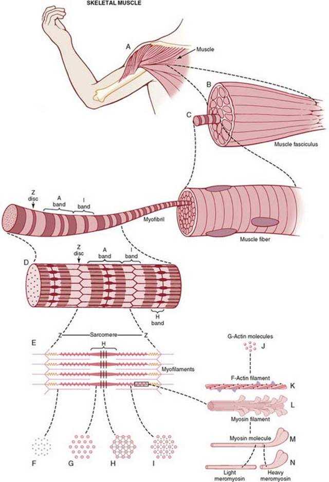
In order to understand the steps of muscle contraction, it is important to first understand the structure of a muscle cell. A muscle cell, also known as a muscle fiber, is made up of bundles of myofibrils, which contain smaller units called sarcomeres. Sarcomeres are the basic functional units of muscles and are responsible for muscle contraction.
The first step of muscle contraction is the activation of the muscle fiber. This occurs when an electrical signal, called an action potential, travels along the motor neuron and reaches the neuromuscular junction. At the neuromuscular junction, the action potential causes the release of a neurotransmitter called acetylcholine. Acetylcholine then binds to receptors on the muscle fiber, activating a series of events that lead to muscle contraction.
Once the muscle fiber is activated, the next step is the sliding filament mechanism. This mechanism involves the interaction between the thin actin filaments and the thick myosin filaments within the sarcomere. When the muscle is at rest, the actin and myosin filaments are not connected. However, when calcium ions are released into the muscle cell, they bind to the protein troponin, causing a conformational change that allows the myosin heads to bind to the actin filaments.
A cycle of events then occurs, known as the cross-bridge cycle, where the myosin heads pull the actin filaments towards the center of the sarcomere. This shortens the sarcomere and ultimately leads to muscle contraction. ATP, the energy molecule of the cell, is needed for the myosin heads to detach from the actin filaments and for the cycle to continue.
In summary, the steps of muscle contraction involve the activation of the muscle fiber, the sliding filament mechanism, and the cross-bridge cycle. These processes occur at the molecular level within the sarcomere and result in the shortening of the muscle fiber, leading to muscle contraction.
Mechanism of Muscle Contraction
Muscle contraction is a complex process that involves the interaction between muscle fibers and nerve signals. It is vital for various biological functions, including movement, posture, and heat production. The mechanism of muscle contraction can be divided into several steps.
The first step in muscle contraction is the release of acetylcholine, a neurotransmitter, from the nerve terminal. The acetylcholine then binds to receptors on the muscle fiber, causing an influx of sodium ions into the muscle cell. This leads to the depolarization of the cell membrane and the generation of an action potential.
Once the action potential is generated, it spreads along the muscle fiber through a network of specialized structures called the T-tubules. This triggers the release of calcium ions from the sarcoplasmic reticulum, a specialized organelle within the muscle cell. The calcium ions then bind to troponin, a protein on the thin filaments, causing a conformational change.
This conformational change in troponin allows the myosin heads, present on the thick filaments, to bind to actin, a protein on the thin filaments. This forms cross-bridges between the thick and thin filaments. The myosin heads then undergo a series of conformational changes, pulling the thin filaments towards the center of the sarcomere, the basic unit of muscle contraction.
This sliding of the filaments results in the shortening of the sarcomere and the overall contraction of the muscle. The duration and force of the contraction are controlled by the frequency and magnitude of the nerve signals received by the muscle fibers. Once the nerve signals stop, the supply of calcium ions is reduced, and the myosin heads detach from actin, leading to the relaxation of the muscle.
In summary, muscle contraction involves the release of acetylcholine, depolarization of the muscle cell, release of calcium ions, binding of myosin heads to actin, sliding of the filaments, and subsequent relaxation of the muscle. This process is highly regulated and allows for the precise control of movement in the body.
The Role of Calcium in Muscle Contraction
Muscle contraction is a complex process that involves the interaction of multiple cellular components and signaling pathways. One of the key players in this process is calcium. Calcium ions play a crucial role in initiating and regulating muscle contraction.
When a muscle is at rest, the concentration of calcium ions is low inside the muscle cells. However, when an action potential travels down the motor neuron and reaches the neuromuscular junction, it triggers the release of a neurotransmitter called acetylcholine. This acetylcholine binds to receptors on the muscle cell membrane, causing the membrane to depolarize and open channels that allow calcium ions to flow into the muscle cell.
Once inside the muscle cell, calcium ions bind to a protein called troponin, which is part of the thin filament of the muscle fiber. This binding causes a conformational change in troponin, which then moves tropomyosin away from the binding sites on the actin filament. This allows myosin, a thick filament, to bind to actin and form cross-bridges. The cross-bridges then undergo a series of conformational changes, resulting in the sliding of the actin and myosin filaments past each other, and ultimately, muscle contraction.
In addition to initiating muscle contraction, the concentration of calcium ions also plays a role in regulating the strength and duration of the contraction. The release of calcium ions from the sarcoplasmic reticulum, a specialized network of tubules within the muscle cell, is tightly controlled by the activity of another protein called the ryanodine receptor. This receptor acts as a calcium release channel, and the level of calcium ions in the muscle cell determines the amount of calcium released and subsequently, the strength of the contraction.
Overall, the role of calcium in muscle contraction is essential. It acts as a trigger for the initiation of contraction and also regulates its strength and duration. Without the presence of calcium ions, muscle contraction would not be possible.
Sliding Filament Theory of Muscle Contraction

The sliding filament theory of muscle contraction is a physiological model that explains how muscles contract and generate force. According to this theory, muscles are made up of overlapping thin and thick filaments. The thin filaments, composed of actin protein, contain binding sites for myosin, while the thick filaments, composed of myosin protein, have small projections called cross-bridges.
When a muscle receives a signal from the nervous system to contract, calcium ions are released, which bind to the regulatory proteins on the actin filaments, allowing the myosin cross-bridges to attach. The myosin cross-bridges then undergo a series of conformational changes, pulling the thin filaments towards the center of the sarcomere. This process is powered by ATP hydrolysis, as the energy released from ATP is used to detach and reattach the myosin heads to the actin filaments.
As the myosin cross-bridges continue to cycle, pulling the thin filaments towards the center of the sarcomere, the sarcomere shortens, resulting in muscle contraction. This sliding filament mechanism allows muscle fibers to generate force and produce movement. The strength and speed of muscle contractions can be modulated by the number of active cross-bridges and the rate at which they cycle.
In summary, the sliding filament theory of muscle contraction explains how muscles generate force and undergo contraction. It involves the interaction between the actin and myosin filaments within the sarcomere, driven by ATP hydrolysis. This process allows muscles to produce the mechanical work required for various physiological activities such as locomotion, posture, and fine motor control. The understanding of this theory is essential for studying and treating muscle-related disorders and optimizing athletic performance.
Regulation of Muscle Contraction

Muscle contraction is a complex process that is regulated by a variety of factors. One key factor in the regulation of muscle contraction is the presence of calcium ions. When a muscle is at rest, the concentration of calcium ions inside the muscle cells is relatively low. However, when a muscle is stimulated to contract, calcium ions are released from storage sites within the muscle cells and bind to proteins called troponin. This binding of calcium to troponin allows another protein, called tropomyosin, to move away from its blocking position on the actin filaments. As a result, myosin heads are able to bind to actin, initiating the sliding of the actin and myosin filaments and ultimately leading to muscle contraction.
Another important regulator of muscle contraction is the neurotransmitter acetylcholine. When a motor neuron stimulates a muscle cell, it releases acetylcholine at the neuromuscular junction, which is the site of communication between the motor neuron and the muscle cell. Acetylcholine binds to receptors on the muscle cell membrane, triggering an electrical impulse that spreads along the membrane and into the muscle cell. This electrical impulse then leads to the release of calcium ions and the subsequent initiation of muscle contraction.
In addition to calcium ions and acetylcholine, the regulation of muscle contraction is also influenced by a number of other factors, including the availability of ATP (the energy source for muscle contraction), the concentration of other ions such as potassium and sodium, and the presence of hormones and other signaling molecules. These various regulatory factors work together to ensure the proper timing and strength of muscle contractions, allowing for efficient movement and coordination of the body.