
Embarking on a rat dissection lab can be an exciting yet daunting experience for students. It is a hands-on opportunity to explore the anatomy of a mammal and gain insight into the complexities of its organ systems. To ensure a successful dissection, it is crucial to have a comprehensive answer key that can guide students through the various steps and provide a deeper understanding of the anatomical structures they will encounter.
This article aims to serve as your ultimate Rat Dissection Lab Answer Key, offering detailed explanations and insights into each step of the dissection process. From the initial exploration of the external anatomy to the intricate examination of the internal organs, you will find essential information and tips that will enhance your learning experience.
With the help of this answer key, students will be able to identify and understand the functions of key anatomical features such as the rat’s skeletal system, respiratory system, digestive system, and circulatory system. By following the step-by-step instructions and referring to the labeled diagrams, students can confidently navigate the dissection process and deepen their knowledge of mammalian anatomy.
Rat Dissection Lab Answer Key
During a rat dissection lab, students are tasked with examining the anatomy and internal organs of a rat. This hands-on activity allows students to apply their knowledge of anatomy and physiology to a real-life specimen. The lab answer key provides a guide for students to refer to while dissecting and identifying the various structures of the rat.
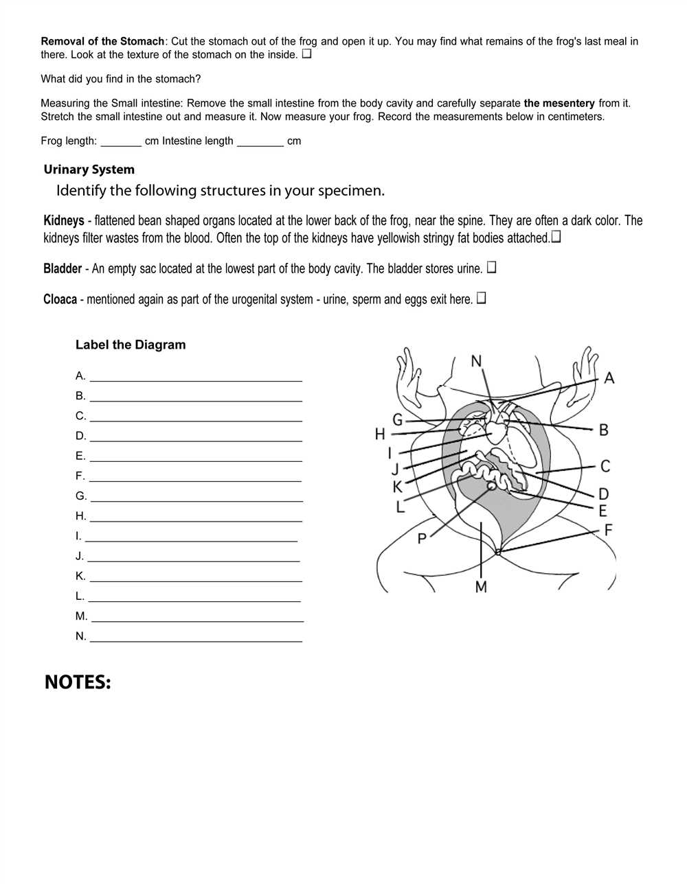
The rat dissection lab answer key typically includes a labeled diagram of the rat, highlighting the major organs and structures that students should be able to identify. This diagram serves as a reference point for students to compare and identify the organs they observe during the dissection. Additionally, the answer key may include written descriptions and explanations of each organ’s function and importance.
Some key structures and organs that students may be asked to identify include:
- Brain: the control center of the nervous system
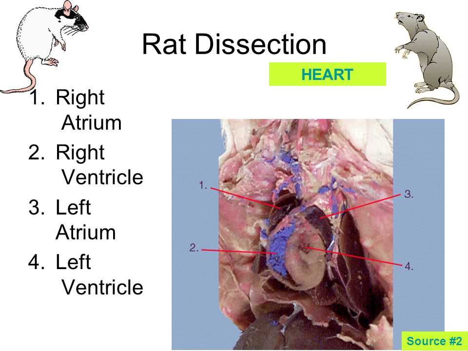
- Heart: responsible for pumping blood throughout the body
- Lungs: involved in the process of respiration
- Liver: plays a role in digestion, metabolism, and detoxification
- Stomach: involved in the breakdown of food
- Intestines: responsible for absorbing nutrients from digested food
- Kidneys: filter waste from the blood and produce urine
By using the rat dissection lab answer key, students can effectively navigate through the dissection process and gain a better understanding of the rat’s anatomy. It also allows for a more structured and organized lab experience, as students have a clear reference point to follow. This lab activity serves as a valuable educational tool for students studying biology and anatomy, providing them with hands-on experience and practical knowledge.
Preparations for Rat Dissection
Before beginning a rat dissection, it is important to have all the necessary materials and tools ready. These include a preserved rat specimen, dissecting tray, gloves, dissecting scissors, forceps, a scalpel, and a dissecting probe. It is also recommended to have a safety goggles and a lab coat to protect yourself during the dissection process.
Once all the materials are gathered, it is important to set up a clean and organized work area. Place the dissecting tray on a stable surface and ensure there is enough lighting for clear visibility. Have a ruler or measuring tape nearby for accurate measurements during the dissection.
Step 1: External Examination
Start the rat dissection by examining the external features of the rat. Observe the overall body structure, the presence of fur, and any external anatomical structures such as the eyes, ears, nose, and tail. Use the ruler or measuring tape to measure the length of the rat from the tip of the nose to the base of the tail. Take note of any abnormalities or differences in coloration.
Step 2: Opening the Body Cavity
To open the body cavity, use the dissecting scissors to make a midline incision from the pubic region to the chin. Be careful not to cut too deep to avoid damaging the underlying organs. Once the incision has been made, use the forceps to gently lift the skin and cut away any excess connective tissue. Take caution not to damage the underlying structures while removing the skin.
Step 3: Internal Examination
With the body cavity open, carefully observe the internal organs. Identify and examine the major organ systems, such as the digestive, respiratory, circulatory, and reproductive systems. Use the dissecting probe to gently explore and manipulate the organs for a better understanding of their structure and function. Take note of any abnormalities or differences observed.
Structure and Function of Rat Anatomy
The rat is a common mammal that is often used in scientific research and dissection labs to study the structure and function of its anatomy. This animal has a complex body structure, with various organ systems that work together to support its life processes. Understanding the anatomy of the rat can provide valuable insights into the similarities and differences between rat and human biology.
One of the key features of the rat’s anatomy is its skeletal system. The rat has a vertebrate skeleton, composed of bones that give the body shape and support. The skeletal system not only provides structural support but also protects vital organs such as the brain, heart, and lungs. Additionally, the rat’s skeleton is responsible for producing blood cells and storing minerals like calcium.
- Skull: The rat’s skull protects the brain and houses the sensory organs, including the eyes, ears, and nose.
- Spine: The rat’s spine, or vertebral column, consists of individual vertebrae that provide flexibility and support.
- Ribs: The rat’s ribs enclose and protect the heart and lungs.
- Limbs: The rat has four limbs, each consisting of a humerus, radius, ulna, and various smaller bones. These limbs allow for movement and coordination.
In addition to the skeletal system, the rat also has a complex digestive system. This system is responsible for breaking down food and absorbing nutrients. The rat has a mouth, which contains teeth for biting and chewing food. From the mouth, the food travels down the esophagus and into the stomach, where it is further broken down by stomach acids. Nutrients are then absorbed in the small intestine, and waste products are eliminated through the rectum and anus.
Overall, studying the structure and function of the rat’s anatomy is essential for understanding mammalian biology. The various organ systems work together to allow the rat to perform essential life processes, and studying these systems can provide insights into human anatomy as well. By dissecting and examining the rat’s anatomy, scientists and students can gain a deeper understanding of mammalian biology and advance their knowledge in the field.
Tools and Techniques for Rat Dissection
When conducting a rat dissection lab, it is important to have the proper tools and techniques in order to successfully navigate the intricacies of the rat’s anatomy. The following are some essential tools that are commonly used during a rat dissection:
- Scalpel: This sharp surgical knife is used to make precise incisions and cuts during the dissection. It is important to handle the scalpel with care and follow proper safety precautions.
- Dissecting scissors: These scissors are designed to cut through tough tissue and small bones. They come in various sizes and shapes to accommodate different dissection needs.
- Forceps: These tweezers-like tools are used to hold and manipulate delicate structures during the dissection. They are particularly useful when dissecting small blood vessels and nerves.
- Dissecting pins: These thin, metal pins are used to hold back flaps of skin or to secure structures in place. They help provide better visibility and access to different parts of the rat’s body.
- Probe: This slender, pointed tool is used to explore and identify structures within the rat’s body. It is helpful for locating and tracing blood vessels, nerves, and organs.
Techniques used during a rat dissection also play a crucial role in obtaining accurate results and minimizing damage to the rat’s tissues. Some common techniques include:
- Initial examination: Before starting the dissection, it is important to thoroughly examine the external anatomy of the rat. This includes observing its size, fur coloration, and any visible abnormalities.
- Systematic dissection: It is recommended to follow a systematic approach when dissecting the rat, starting from the external structures and gradually moving deeper into the body. This helps ensure that all structures are properly examined and dissected in a logical order.
- Proper handling and cutting: It is essential to handle the rat with care and to make precise, clean cuts during the dissection. This helps avoid unnecessary damage to the tissues and organs being examined.
- Recording observations: As you progress through the dissection, it is important to document your observations and findings. This may include noting the appearance, size, and location of structures, as well as any abnormalities or variations.
- Dispose of materials properly: After completing the dissection, it is important to properly dispose of all materials used, including the rat carcass. Follow proper guidelines and regulations for biohazardous waste disposal to ensure safety.
By using the appropriate tools and techniques, a rat dissection lab can be conducted effectively and with precision. This allows for a thorough exploration of the rat’s anatomy and provides valuable insights for further study in the field of biology.
Step-by-Step Guide to Rat Dissection
Dissecting a rat can be an instructive and eye-opening experience for students studying anatomy or biology. It provides an opportunity to understand the internal structures and functions of a mammal and gain hands-on experience in a laboratory setting. Here is a step-by-step guide to rat dissection:
1. Preparation
Begin by gathering all the necessary materials, including a preserved rat, dissecting tools (such as scalpels and scissors), gloves, goggles, and a dissection tray. Make sure to review the safety precautions and guidelines before starting the dissection.
2. External Examination
Place the rat on the dissection tray and observe its external features. Note the size, fur color, and any visible abnormalities. Pay attention to the different anatomical regions such as the head, neck, limbs, and tail.
3. Incision
Use a scalpel or scissors to make a midline incision from the neck to the tail. Be careful not to cut too deep to avoid damaging internal structures. Carefully separate the skin and muscles from the body wall and pin them back to expose the internal organs.
4. Organ Identification
Begin by identifying and examining the major organs, such as the heart, lungs, liver, kidneys, and intestines. Observe their size, shape, color, and texture. Take note of any differences compared to human anatomy.
5. Systematic Dissection
Continue the dissection by systematically examining each organ system. Use a scalpel or scissors to carefully dissect and explore the nervous, digestive, respiratory, circulatory, and reproductive systems. Take your time to locate and understand each organ within these systems.
6. Documentation
As you progress through the dissection, take notes and draw diagrams to document your findings. Label the structures and organs you have identified, and make observations about their functions and connections to other systems.
7. Disposal
Once you have completed the dissection, clean up the workspace and properly dispose of the rat and any dissected tissues. Follow the guidelines provided by your instructor or institution for disposal.
Remember to work carefully and respectfully throughout the dissection process. Take the opportunity to ask questions, seek clarification, and discuss your findings with your classmates and instructor. Rat dissection can be a valuable learning experience that enhances your understanding of mammalian anatomy and physiology.
Observation and Analysis of Rat Organs
During the rat dissection lab, we carefully observed and analyzed the various organs of the rat. The dissection allowed us to gain a deeper understanding of the structure and function of these organs and how they contribute to the overall physiology of the rat.
The digestive system: The rat’s digestive system consists of several organs, including the mouth, esophagus, stomach, small intestine, and large intestine. We observed that the rat’s teeth were well-adapted for chewing and grinding food, and the stomach was muscular and capable of breaking down the food further. The small intestine was lined with finger-like projections called villi, which increased the surface area for nutrient absorption. The large intestine was responsible for reabsorbing water and forming feces.
The respiratory system: We examined the rat’s lungs, trachea, and diaphragm as part of the respiratory system. The lungs were soft and spongy and provided a large surface area for gas exchange. The trachea was a rigid tube that allowed air to pass into and out of the lungs. The diaphragm, a dome-shaped muscle, played a crucial role in breathing by contracting and relaxing to expand and contract the lungs.
The circulatory system: The rat’s circulatory system included the heart, blood vessels, and blood. We observed that the heart was a four-chambered organ responsible for pumping oxygenated blood to the body and deoxygenated blood to the lungs. The blood vessels, including arteries, veins, and capillaries, facilitated the transportation of oxygen and nutrients to the body’s cells. We also examined the blood, noting its red color due to the presence of hemoglobin and its role in carrying oxygen.
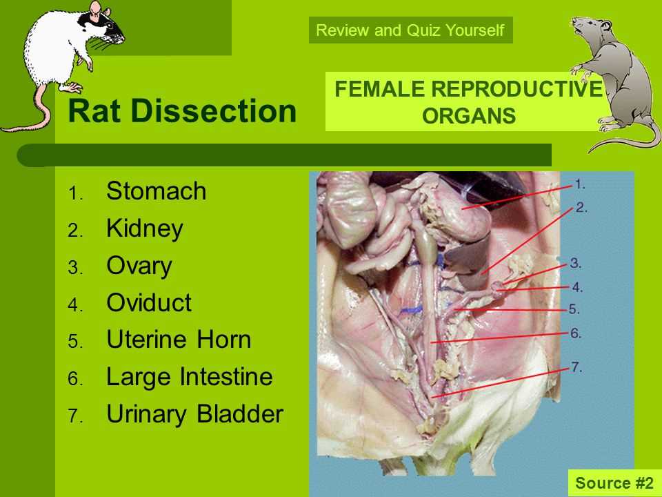
The urinary system: The rat’s urinary system comprised the kidneys, ureters, bladder, and urethra. We observed that the kidneys were bean-shaped organs responsible for filtering waste products from the blood and producing urine. The urine then traveled through the ureters to the bladder, where it was stored until eliminated through the urethra. The urinary system played a vital role in maintaining the rat’s homeostasis by regulating the balance of water and electrolytes in the body.
Overall, the observation and analysis of the rat’s organs during the dissection lab provided us with valuable insights into the complexity and functionality of these organ systems. It reinforced the interconnectedness of the various systems in maintaining the rat’s overall health and survival.