
The spinal cord is a crucial component of the central nervous system, responsible for transmitting signals between the brain and the rest of the body. It is a long, thin, tubular bundle of nerves that extends from the base of the brain to the lower back. Understanding the workings of the spinal cord and its relationship with the reflex act is essential to comprehend the complexities of the human nervous system.
One way to gain a deeper understanding of the spinal cord and reflex act is through a worksheet that presents various scenarios and questions. This worksheet tests our knowledge of the spinal cord’s anatomy, functions, and reflex actions. By providing answers to the worksheet, we can enhance our comprehension of these intricate systems and their interplay in the human body.
One of the questions on the worksheet might involve identifying the main components of the spinal cord. In this case, we would need to highlight the gray matter, white matter, and the central canal. The gray matter consists of nerve cell bodies, while the white matter contains nerve fibers. The central canal is a narrow channel filled with cerebrospinal fluid that runs the length of the spinal cord.
Overview of the Spinal Cord and Reflex Act
The spinal cord is a vital part of the central nervous system that runs down the back and acts as a communication pathway between the brain and the body. It is responsible for transmitting sensory information from the body to the brain, as well as sending motor commands from the brain to the muscles and organs.
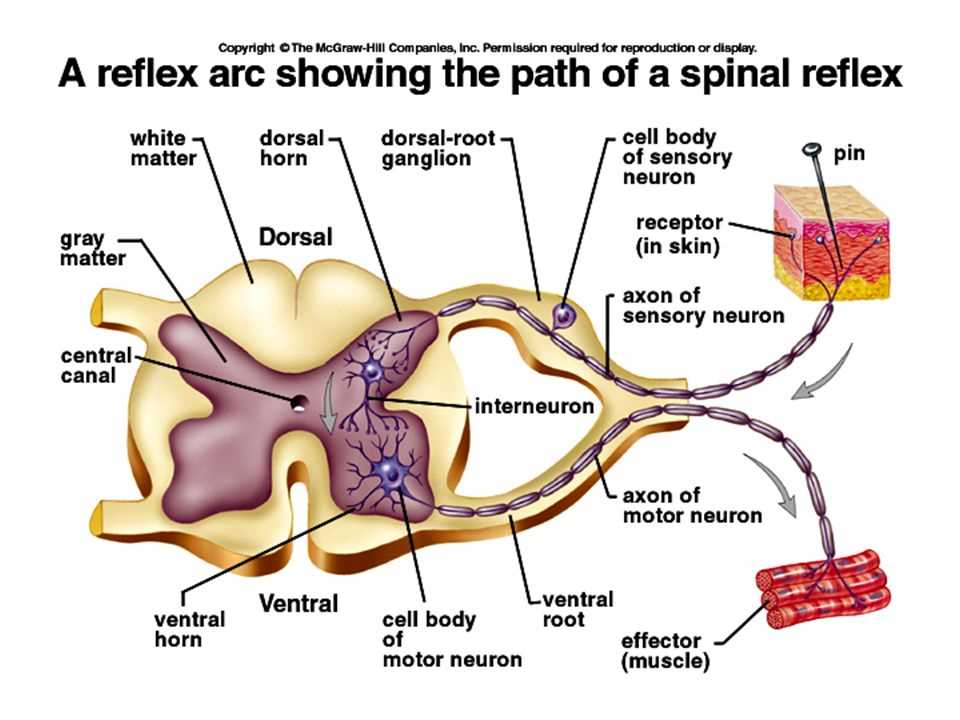
One of the important functions of the spinal cord is its involvement in the reflex act. A reflex act is an automatic and involuntary response to a stimulus, which occurs without conscious thought. The reflex arc, which includes the spinal cord, is responsible for coordinating these quick responses to potentially dangerous or harmful stimuli.
The process of the reflex act involves several steps:
- Sensory receptors in the body detect a stimulus, such as heat or pain.
- The sensory neurons transmit the sensory information to the spinal cord.
- The information is then relayed to the motor neurons in the spinal cord.
- The motor neurons then send signals to the appropriate muscles to produce a rapid response, such as moving away from the source of pain.
One of the key advantages of the reflex act is its speed. Because the sensory information is processed in the spinal cord rather than having to travel all the way to the brain, reflex responses can occur within milliseconds. This rapid response time can help protect the body from potential harm.
Overall, the spinal cord and reflex act play crucial roles in both sensory perception and motor control. Their efficient and quick communication allows for swift reactions to external stimuli, helping to ensure the well-being and survival of the individual.
The Role of the Spinal Cord in the Nervous System
The spinal cord is a crucial component of the nervous system, playing a vital role in the transmission of signals between the brain and the rest of the body. It serves as a highway for communication, allowing sensory information to be relayed from the peripheral nervous system to the brain, and motor signals to be sent from the brain to the muscles and organs.
One of the key functions of the spinal cord is to coordinate reflex actions. Reflexes are rapid, involuntary responses to specific stimuli that help protect the body and maintain homeostasis. Examples of reflex actions include pulling your hand away from a hot object or automatically blinking when an object approaches your eye. The spinal cord plays a crucial role in these reflexes by integrating and processing sensory information and triggering an immediate motor response without the involvement of the brain.
The spinal cord is made up of a complex network of neurons, divided into different regions responsible for specific functions. The grey matter in the center of the cord contains cell bodies of neurons, while the white matter on the outside consists of nerve fibers that transmit signals up and down the cord. Sensory neurons enter the spinal cord through the dorsal (posterior) root, while motor neurons exit through the ventral (anterior) root.
Overall, the spinal cord serves as an essential link between the brain and the rest of the body, allowing for the coordination of complex voluntary movements, as well as the initiation of fast reflex actions. It is a remarkable structure that plays a fundamental role in our daily activities and helps ensure our survival and well-being.
Understanding Reflex Acts and Their Importance
Reflex acts are automatic responses that occur in the body without conscious thought. They are essential for our daily functioning and play a crucial role in protecting us from harm. By understanding reflex acts, we can gain insight into the complexity of our nervous system and appreciate the incredible mechanisms that allow our bodies to respond quickly and effectively to potential dangers.
The Spinal Cord: The spinal cord is a significant component in the pathway of reflex acts. It acts as a relay station, receiving sensory information from various parts of the body and sending motor commands back to initiate the appropriate response. This quick transfer of signals enables reflex actions to occur rapidly, often before we are even consciously aware of the stimulus.
The Importance of Reflex Acts: Reflex acts are vital for our survival and well-being. They serve as our body’s first line of defense against potentially harmful stimuli, allowing us to react swiftly and instinctively. For example, the withdrawal reflex helps us remove our hand from a hot object without conscious thought, preventing severe burns. Similarly, the patellar reflex helps maintain our balance and stability by ensuring that our knee-jerk reaction remains intact.
Reflex vs. Voluntary Actions: While reflex acts are automatic and involuntary, voluntary actions involve conscious decision-making. Reflex acts are quicker and do not require the involvement of higher brain centers, making them fundamental for our immediate response to certain situations. Understanding the difference between reflex and voluntary actions helps us appreciate the intricate coordination between our sensory organs, spinal cord, and brain.
Reflex Acts and Neurological Disorders: Studying reflex acts also provides valuable insights into various neurological disorders. For instance, an absence or abnormality in specific reflexes may indicate damage or dysfunction in certain areas of the nervous system. These reflex tests help healthcare professionals diagnose and monitor conditions such as spinal cord injuries, multiple sclerosis, and certain neurodegenerative diseases.
Overall, understanding reflex acts and their importance allows us to appreciate the remarkable capabilities of our nervous system. They showcase the intricate coordination between our sensory organs, spinal cord, and brain and play a critical role in maintaining our safety and well-being.
Anatomy of the Spinal Cord

The spinal cord is a long, cylindrical structure that extends from the base of the skull to the lower back. It is an integral part of the central nervous system and is responsible for transmitting signals between the brain and the rest of the body. The spinal cord is protected by the vertebral column, or backbone, which consists of individual vertebrae stacked on top of each other.
The spinal cord is composed of gray matter and white matter. The gray matter is located in the central region of the spinal cord and is shaped like a butterfly. It contains cell bodies and dendrites of nerve cells, as well as synapses where nerve impulses are transmitted. The white matter surrounds the gray matter and is made up of myelinated nerve fibers that carry nerve impulses up and down the spinal cord.
The spinal cord is organized into segments, each of which corresponds to a specific region of the body. There are a total of 31 segments, and each segment has a pair of spinal nerves that emerge from it. These spinal nerves are responsible for transmitting sensory information from the body to the brain and motor signals from the brain to the muscles and glands.
In addition to its role in transmitting signals, the spinal cord is also involved in coordinating reflex actions. Reflexes are rapid, involuntary responses to stimuli that do not involve the brain. They are mediated by circuits within the spinal cord called reflex arcs. Reflex arcs allow for quick reactions to potential danger or harm, such as pulling your hand away from a hot surface without having to consciously think about it.
The anatomy of the spinal cord is complex and plays a crucial role in the functioning of the nervous system. Understanding its structure and function is essential for diagnosing and treating conditions that affect the spinal cord, such as spinal cord injuries and disorders.
Anatomy of the Spinal Cord:
- Long, cylindrical structure
- Extends from the base of the skull to the lower back
- Protected by the vertebral column
- Composed of gray matter and white matter
- Gray matter contains cell bodies and synapses
- White matter consists of myelinated nerve fibers
- Organized into segments, each with a pair of spinal nerves
Structure and Components of the Spinal Cord
The spinal cord is a long, cylindrical structure that runs through the vertebral column and serves as a crucial part of the central nervous system (CNS). It is made up of millions of nerve fibers that transmit electrical signals between the brain and peripheral nervous system.
The spinal cord can be divided into different regions, each with specific functions. The cervical region, located in the neck, is responsible for transmitting nerve signals to and from the upper limbs, as well as controlling the muscles and sensory input of the neck and upper back. The thoracic region, which is in the middle of the spine, plays a role in transmitting signals to and from the thorax and abdomen. The lumbar region, located in the lower back, is responsible for transmitting signals to and from the lower limbs and controlling the muscles and sensory input of the lower back and legs. Finally, the sacral region is involved in transmitting signals to and from the pelvic organs and lower limbs.
The spinal cord has different components that work together to carry out its functions. The gray matter, which forms the inner region of the spinal cord, consists of nerve cell bodies, dendrites, and unmyelinated axons. It processes and integrates sensory information and controls voluntary motor movements. The white matter, which surrounds the gray matter, is made up of myelinated axons that transmit signals up and down the spinal cord. It forms tracts that connect different parts of the central nervous system.
Overall, the spinal cord is a complex structure that plays a vital role in the functioning of the human body. Its structure and components allow for the transmission of information between the brain and the rest of the body, as well as the coordination of movements and sensory input.
Functions of Different Segments of the Spinal Cord
The spinal cord, which is part of the central nervous system, plays a crucial role in transmitting information between the brain and the rest of the body. It is divided into different segments, each with its own specific functions. These segments include the cervical, thoracic, lumbar, sacral, and coccygeal regions.
Cervical Segment:
The cervical segment of the spinal cord is located in the neck region and is responsible for controlling the movement and sensation of the upper limbs, neck, and shoulders. It also contains the nerves that control the diaphragm, the main muscle involved in breathing.
Thoracic Segment:
The thoracic segment of the spinal cord is located in the chest region and is primarily responsible for transmitting sensory information from the trunk and controlling the movement and sensation of the trunk muscles. It also helps regulate the functioning of the internal organs such as the heart, lungs, and digestive system.
Lumbar Segment:
The lumbar segment of the spinal cord is located in the lower back and is responsible for controlling the movement and sensation of the lower limbs. It plays a key role in maintaining balance and coordinating movements such as walking and running.
Sacral and Coccygeal Segments:
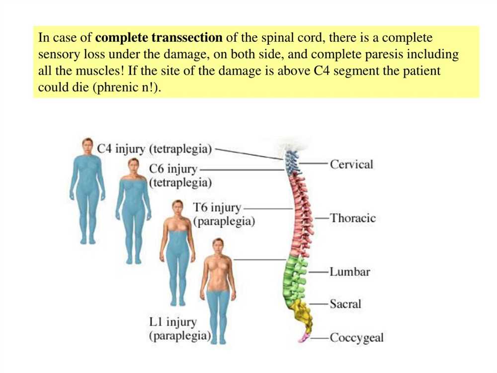
The sacral and coccygeal segments of the spinal cord are located in the pelvic region and are involved in controlling the functions of the bladder, bowel, and sexual organs. These segments also play a role in transmitting sensory information from the skin and muscles in the lower pelvic region.
In summary, each segment of the spinal cord serves specific functions related to movement, sensation, and control of various body parts and organ systems. Understanding these functions is important for identifying and diagnosing disorders or injuries that may affect specific segments of the spinal cord.
Pathways of Sensory and Motor Information
The pathways through which sensory and motor information travel play a crucial role in our ability to perceive and respond to the world around us. These pathways consist of a complex network of nerves that transmit signals from our sensory receptors to our brain, and from our brain to our muscles and glands.
Sensory pathways are responsible for carrying information about our external environment (such as touch, temperature, and pain) as well as our internal environment (such as the position of our limbs and the status of our organs). They begin with sensory receptors located in various parts of our body, which detect and convert different types of stimuli into electrical signals. These signals then travel along sensory neurons, passing through different levels of the spinal cord and brain before reaching their final destination in the sensory cortex, where they are interpreted and consciously perceived.
Motor pathways, on the other hand, are responsible for carrying signals from our brain to our muscles and glands, enabling us to perform voluntary movements and control bodily functions. They begin with motor cortex, which is located in the frontal lobe of the brain. From there, signals are transmitted through the spinal cord, where they are relayed to the appropriate motor neurons that innervate specific muscles. These signals trigger a series of events that result in muscle contraction or glandular secretion, allowing us to carry out various actions, from walking and talking to digestion and perspiration.
It is important to note that sensory and motor pathways are not independent, but rather interconnected. This allows for the seamless integration of sensory information and motor commands, enabling us to respond to sensory stimuli in a coordinated and appropriate manner. For example, when we touch something hot, sensory information is quickly transmitted to the brain, which then sends a signal through the motor pathways to retract our hand, preventing injury. This reflexive action is an example of the remarkable efficiency and speed of these pathways, which are crucial for our survival and daily functioning.