
Understanding the intricate workings of a synapse, the junction between two nerve cells, has long been a subject of fascination for scientists. This microscopic structure plays a crucial role in transmitting information throughout the nervous system, enabling us to think, feel, and move. In recent years, researchers have made significant strides in unraveling the mysteries of synapses, shedding light on the complex mechanisms behind their formation, function, and communication.
One question that has captivated neuroscientists is how synapses are structured and organized. The anatomy of a synapse holds the key to understanding its function. Within a synapse, there are numerous components, each with a specific role to play. At the heart of the synapse is the synaptic cleft, a narrow gap between the presynaptic and postsynaptic neurons. Spanning this gap are proteins called neurotransmitter receptors, which serve as the connecting link between the two neurons. These receptors are responsible for receiving chemical signals, or neurotransmitters, released by the presynaptic neuron and relaying them to the postsynaptic neuron.
Another crucial element within a synapse is the presynaptic terminal, where neurotransmitters are stored and released. This terminal contains tiny sacs called synaptic vesicles, which house the neurotransmitters until they are ready to be released into the synaptic cleft. When an electrical signal reaches the presynaptic neuron, these vesicles fuse with the presynaptic membrane, releasing the neurotransmitters into the synaptic cleft. This process, known as exocytosis, is the key to transmitting information from one neuron to another.
The Anatomy of a Synapse Answers
A synapse is a small, microscopic structure in the nervous system that allows neurons to communicate with each other. It is the site where electrical impulses are transmitted from one neuron to another, enabling information to be transferred throughout the brain and spinal cord. The anatomy of a synapse consists of several key components that work together to facilitate this communication.
At the center of a synapse is the synaptic cleft, a small gap between the axon terminal of one neuron and the dendrites or cell body of another neuron. The synaptic cleft is filled with a fluid-like substance called extracellular fluid, which helps to transmit signals across the synapse. Surrounding the synapse are glial cells, which provide support and insulation to the neurons.
The pre-synaptic neuron, or the neuron sending the signal, is connected to the post-synaptic neuron, or the neuron receiving the signal, through specialized structures called synaptic vesicles. These vesicles contain neurotransmitters, which are chemical messengers that are released into the synaptic cleft when an electrical impulse reaches the axon terminal.
When a neurotransmitter is released into the synaptic cleft, it binds to specific receptors on the post-synaptic neuron, initiating a series of chemical reactions. This binding triggers an electrical impulse in the post-synaptic neuron, allowing the signal to be transmitted from one neuron to another. Once the signal is transmitted, the neurotransmitter is either reabsorbed by the pre-synaptic neuron or broken down by enzymes in the synaptic cleft.
In summary, the anatomy of a synapse includes the synaptic cleft, glial cells, synaptic vesicles, neurotransmitters, and receptors. These components work together to facilitate the transmission of electrical impulses between neurons, allowing for communication and information processing in the nervous system.
What is a Synapse?
A synapse is a specialized connection between two nerve cells, or neurons, in the brain. It is the fundamental unit of communication in the nervous system, allowing for the transmission of information between neurons. Synapses are crucial for the functioning of the brain and are involved in processes such as learning, memory, and perception.
At a synapse, there are typically two main components: the presynaptic neuron, which sends the signal, and the postsynaptic neuron, which receives the signal. These cells are separated by a small gap known as the synaptic cleft. The synapse functions through a process called neurotransmission, where chemical signals, or neurotransmitters, are released by the presynaptic neuron and bind to receptors on the postsynaptic neuron, allowing for the transfer of information.
Key Points:
- Synapses are specialized connections between neurons in the brain.
- They allow for the transmission of information between neurons through neurotransmitters.
- Synapses are essential for processes such as learning, memory, and perception.
Overall, synapses play a vital role in the functioning of the nervous system and are crucial for various cognitive processes. Understanding the anatomy and function of synapses is essential for gaining insight into how the brain works and how it processes information.
Structure of a Synapse
A synapse is a specialized junction between two neurons, where signals are transmitted from one neuron to another. It is the fundamental functional unit of the nervous system, allowing for the transmission of information and the integration of neural circuits. The structure of a synapse is highly complex and consists of several key components.
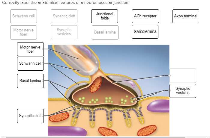
Presynaptic Terminal: The presynaptic terminal, also known as the axon terminal, is located at the end of the presynaptic neuron. It contains vesicles filled with neurotransmitters, which are released into the synaptic cleft.
Synaptic Cleft: The synaptic cleft is a tiny gap between the presynaptic terminal and the postsynaptic membrane of the receiving neuron. The neurotransmitters released from the presynaptic terminal diffuse across this gap to the postsynaptic membrane.
Postsynaptic Membrane: The postsynaptic membrane is the membrane of the receiving neuron, opposite to the presynaptic terminal. It contains receptors that bind to the neurotransmitters released by the presynaptic neuron.
Neurotransmitters: Neurotransmitters are chemical messengers that transmit signals across the synaptic cleft. They are stored in vesicles within the presynaptic terminal and are released upon the arrival of an action potential.
Receptors: Receptors are proteins located on the postsynaptic membrane that bind to specific neurotransmitters. When neurotransmitters bind to their corresponding receptors, they can either initiate or inhibit a response in the receiving neuron.
Synaptic Vesicles: Synaptic vesicles are small membrane-bound compartments within the presynaptic terminal that store neurotransmitters until they are released into the synaptic cleft.
Synaptic Transmission: Synaptic transmission refers to the process of signal transmission across the synapse. It involves the release of neurotransmitters from the presynaptic terminal, the diffusion of neurotransmitters across the synaptic cleft, and the binding of neurotransmitters to receptors on the postsynaptic membrane.
Overall, the structure of a synapse is crucial for the proper functioning of the nervous system. It allows for the efficient transmission and integration of signals, enabling complex processes such as learning, memory, and perception.
Pre-Synaptic Cell
The pre-synaptic cell is an important component of the synapse, playing a crucial role in the transmission of signals between neurons. It is the cell that releases neurotransmitters into the synaptic cleft, which then bind to receptors on the post-synaptic cell to initiate a response. The pre-synaptic cell can be either a neuron or a specialized cell in the nervous system, such as a motor neuron or a sensory neuron.
In the pre-synaptic cell, the process of neurotransmitter release is carefully regulated. When an action potential reaches the end of the pre-synaptic neuron, it triggers the opening of voltage-gated calcium channels. This allows calcium ions to enter the cell, which in turn triggers the release of neurotransmitters from specialized vesicles called synaptic vesicles. The neurotransmitters are then released into the synaptic cleft through a process called exocytosis.
The pre-synaptic cell also contains numerous other components that are essential for efficient neurotransmitter release and synaptic function. These include mitochondria, which provide energy for the cell, and a network of proteins and cytoskeletal elements that help maintain the structure and stability of the synapse. Additionally, the pre-synaptic cell may contain specialized proteins and enzymes that regulate the recycling and reuptake of neurotransmitters, ensuring that the synaptic cleft is cleared of excess neurotransmitters after each transmission.
Overall, the pre-synaptic cell is a key player in synaptic signaling, responsible for the release of neurotransmitters and the regulation of synaptic function. Its intricate structure and complex molecular machinery allow for precise and efficient communication between neurons, enabling the proper functioning of the nervous system.
Post-Synaptic Cell
The post-synaptic cell is a crucial component of the synapse, where the transmission of signals between neurons occurs. It is a specialized cell that receives information from the pre-synaptic neuron via the synaptic cleft. The structure and function of the post-synaptic cell play a vital role in determining the strength and effectiveness of the synaptic transmission.
The post-synaptic cell contains receptors that are specific to the neurotransmitters released by the pre-synaptic neuron. These receptors are embedded in the membrane of the post-synaptic cell and are responsible for detecting the presence of neurotransmitters in the synaptic cleft. Once a neurotransmitter binds to its corresponding receptor, it initiates a series of biochemical reactions that lead to changes in the post-synaptic cell’s membrane potential.
The activation of the receptors triggers the opening or closing of ion channels, allowing ions, such as sodium, potassium, and calcium, to flow into or out of the post-synaptic cell. This influx or efflux of ions generates an electrical charge, known as the post-synaptic potential, which determines whether the post-synaptic cell will fire an action potential and transmit the signal to subsequent neurons.
In addition to ion channels, the post-synaptic cell also contains various proteins and enzymes that contribute to the processing and integration of the incoming signals. These proteins and enzymes regulate the strength and duration of the post-synaptic potential, ensuring precise and reliable communication between neurons.
In summary, the post-synaptic cell is a specialized cell that receives signals from the pre-synaptic neuron and converts them into electrical impulses. It contains receptors that detect neurotransmitters, ion channels that generate post-synaptic potentials, and various proteins and enzymes that regulate synaptic transmission. Understanding the anatomy and physiology of the post-synaptic cell is crucial for unraveling the complexities of neuronal communication and the functioning of the nervous system.
Neurotransmitters
In the context of the anatomy of a synapse, neurotransmitters play a crucial role in the communication between neurons. These chemical messengers are synthesized and stored within the presynaptic neuron, ready to be released into the synaptic cleft upon the arrival of an action potential.
Key facts about neurotransmitters:
- Neurotransmitters can be excitatory or inhibitory, meaning they either increase or decrease the likelihood of an action potential being generated in the postsynaptic neuron.
- Some well-known neurotransmitters include serotonin, dopamine, acetylcholine, and gamma-aminobutyric acid (GABA).
- Each neurotransmitter has its own specific receptors on the postsynaptic neuron, allowing for precise signaling and control.
Excitatory and Inhibitory Neurotransmitters
Excitatory neurotransmitters, such as glutamate, have the ability to depolarize the postsynaptic membrane, bringing it closer to its threshold for an action potential. These neurotransmitters promote neuron firing and facilitate the transmission of signals.
In contrast, inhibitory neurotransmitters, like GABA, hyperpolarize the postsynaptic membrane, making it less likely for an action potential to occur. These neurotransmitters dampen excessive neuronal activity and help maintain a balance within the neural circuitry.
Specific Receptors
Neurotransmitters interact with specific receptors located on the postsynaptic neuron. These receptors can be ion channels or G-protein-coupled receptors, and their activation by neurotransmitters initiates a cascade of intracellular events that ultimately influence the behavior of the postsynaptic neuron.
The specificity between neurotransmitters and their receptors allows for precise communication within the nervous system. Different neurotransmitters bind to different receptor subtypes, and the effects they produce vary accordingly. This complexity contributes to the diversity of neuronal signaling and the ability of the brain to process and integrate information from various sources.
Synaptic Transmission
Synaptic transmission is the process by which information is transmitted between neurons at a synapse. It is a complex and highly regulated process that involves a series of molecular events.
When an action potential reaches the presynaptic terminal, it triggers the release of neurotransmitters into the synapse. These neurotransmitters are stored in small vesicles in the presynaptic terminal. The action potential causes these vesicles to fuse with the presynaptic membrane, releasing their contents into the synapse.
The released neurotransmitters then bind to receptors on the postsynaptic membrane, initiating a series of events that lead to the generation of a new action potential in the postsynaptic neuron. This process is called synaptic transmission. The neurotransmitters can bind to different types of receptors, which can have excitatory or inhibitory effects on the postsynaptic neuron.
After the neurotransmitters have performed their function, they can be broken down or taken back up by the presynaptic neuron in a process called reuptake. This helps to terminate the signaling process and allow for new signals to be transmitted.
In summary, synaptic transmission is a crucial process in the communication between neurons. It involves the release of neurotransmitters, their binding to receptors, and the generation of new action potentials in the postsynaptic neuron. Understanding the details of synaptic transmission is important for unraveling the complexities of neural circuits and for developing new treatments for neurological disorders.