
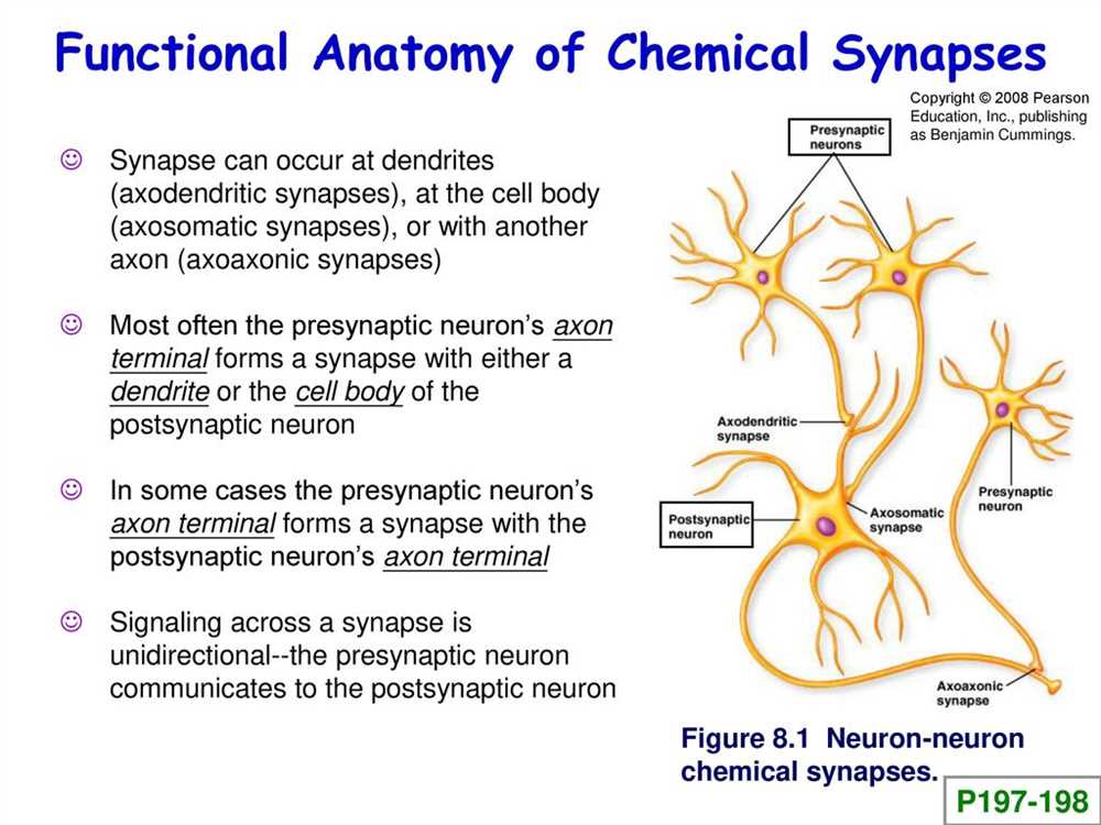
The human brain is an incredibly complex and fascinating organ, consisting of billions of nerve cells called neurons. These neurons communicate with each other through specialized connections called synapses. Understanding the anatomy of a synapse is crucial in unraveling the mechanisms behind brain function and how it relates to various disorders and diseases.
When coloring a synapse, it is important to focus on the key components that make up this intricate structure. One of the most prominent parts is the presynaptic terminal, which contains vesicles filled with neurotransmitters. These neurotransmitters are released into the synaptic cleft, the small gap between the pre- and postsynaptic neurons, and play a crucial role in transmitting signals across the synapse.
Another important component of a synapse is the postsynaptic membrane, which contains receptors that bind to the neurotransmitters and initiate a response in the receiving neuron. These receptors can be excitatory, promoting the generation of an electrical signal in the postsynaptic neuron, or inhibitory, preventing the generation of such a signal. The coloration of these receptors can help visualize the different types of neurotransmitters and their effects on neural communication.
Furthermore, the coloring of the synapse should also include the presence of auxiliary structures, such as astrocytes and glial cells, which provide support and nourishment to the neurons. These cells are essential for maintaining the health and proper functioning of the synapse. By highlighting these auxiliary structures, we can better understand their role in synapse formation, maintenance, and plasticity.
The Anatomy of a Synapse Coloring Answers
Understanding the anatomy of a synapse is crucial for studying the functioning of the nervous system. One way to visualize and learn about the different structures involved is through coloring activities. By coloring different components of a synapse, students can enhance their understanding of this complex process.
Here are the answers for the anatomy of a synapse coloring activity:
- Synaptic Terminal: Color the synaptic terminal orange. This is the end of the axon where neurotransmitters are released.
- Presynaptic Membrane: Color the presynaptic membrane blue. This is the membrane of the synaptic terminal that faces the synaptic cleft.
- Postsynaptic Membrane: Color the postsynaptic membrane yellow. This is the membrane of the receiving neuron that is opposite the presynaptic membrane.
- Synaptic Cleft: Color the synaptic cleft green. This is the small gap between the presynaptic and postsynaptic membranes.
- Neurotransmitters: Color the neurotransmitters red. These are the chemical messengers that are released into the synaptic cleft.
- Receptor Proteins: Color the receptor proteins purple. These proteins are located on the postsynaptic membrane and bind to neurotransmitters.
By engaging in coloring activities like this, students can reinforce their understanding of the anatomical structures involved in synaptic transmission. This visual approach allows for a more interactive and hands-on learning experience, making it easier to remember and grasp the complexities of the synapse.
What is a Synapse?

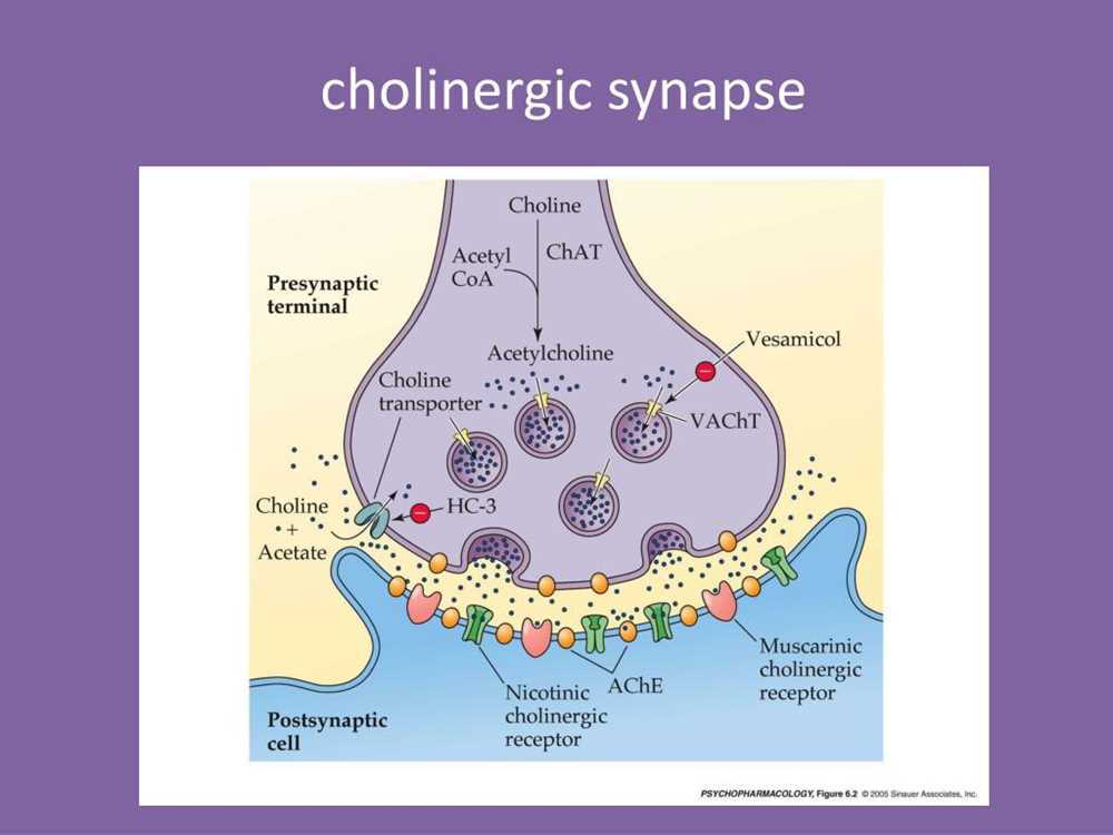
A synapse is a junction between two nerve cells, where signals are transmitted from one cell to another. It is the fundamental unit of communication in the nervous system and plays a crucial role in allowing the transmission of electrical and chemical signals between neurons. A synapse consists of three main components: the presynaptic terminal, the synaptic cleft, and the postsynaptic terminal.
The presynaptic terminal, also known as the axon terminal, is the end of the axon of the sending neuron. It contains vesicles that house neurotransmitters, which are chemical messengers that transmit signals across the synapse. When an action potential reaches the presynaptic terminal, it triggers the release of these neurotransmitters into the synaptic cleft.
The synaptic cleft is the narrow gap between the presynaptic and postsynaptic terminals. It acts as a physical barrier, preventing direct electrical transmission between neurons. Instead, communication occurs through the release and binding of neurotransmitters that diffuse across the cleft.
The postsynaptic terminal, also known as the dendritic spine, is the specialized region of the receiving neuron that contains receptor molecules. These receptors bind to the neurotransmitters released by the presynaptic terminal, initiating a series of chemical and electrical events that result in the generation of an electrical signal in the postsynaptic neuron. This signal can then propagate along the neuron, allowing for the transmission of information within the nervous system.
In conclusion, a synapse is a crucial component of the nervous system that enables communication between neurons. It consists of the presynaptic terminal, synaptic cleft, and postsynaptic terminal, working together to transmit electrical and chemical signals. Understanding the anatomy and function of synapses is essential for understanding the complexities of the nervous system and the processes underlying brain function.
The Structure of a Synapse
A synapse is a structure that allows information to be transmitted from one neuron to another in the form of chemical signals. It consists of three main components: the presynaptic terminal, the synaptic cleft, and the postsynaptic membrane.
The presynaptic terminal is the end of the axon of the releasing (presynaptic) neuron. It contains specialized structures called synaptic vesicles, which store neurotransmitters. When an action potential reaches the presynaptic terminal, it triggers the release of neurotransmitters into the synaptic cleft.
The synaptic cleft is the narrow gap between the presynaptic terminal and the postsynaptic membrane. It is filled with a gel-like substance called extracellular matrix. The neurotransmitters released from the presynaptic terminal diffuse across the synaptic cleft and bind to receptors on the postsynaptic membrane.
The postsynaptic membrane is the membrane of the receiving (postsynaptic) neuron. It contains receptors that are specific to certain neurotransmitters. When neurotransmitters bind to the receptors on the postsynaptic membrane, they can either excite or inhibit the postsynaptic neuron, depending on the type of neurotransmitter and the specific receptor.
- The structure of a synapse allows for the precise and selective transmission of information between neurons.
- The presynaptic terminal releases neurotransmitters into the synaptic cleft.
- The synaptic cleft is the gap between the presynaptic terminal and the postsynaptic membrane.
- The postsynaptic membrane contains receptors that bind to neurotransmitters.
- Neurotransmitters can either excite or inhibit the postsynaptic neuron.
The Pre-Synaptic Neuron
The pre-synaptic neuron plays a vital role in the transmission of signals in the nervous system. It is responsible for initiating and releasing neurotransmitters into the synaptic cleft, where they can bind to receptors on the post-synaptic neuron. This process is crucial for communication between neurons and allows for the transmission of information throughout the body.
The pre-synaptic neuron contains several important components that enable the release of neurotransmitters. One of these components is the axon terminal, which is located at the end of the neuron’s axon. The axon terminal contains synaptic vesicles, which store the neurotransmitters until they are ready to be released. When an action potential reaches the axon terminal, it triggers the opening of voltage-gated calcium channels, allowing calcium ions to enter the neuron.
The entry of calcium ions into the neuron triggers the fusion of the synaptic vesicles with the pre-synaptic membrane. This fusion process releases the neurotransmitters into the synaptic cleft. The neurotransmitters then diffuse across the synaptic cleft and bind to receptors on the post-synaptic neuron, initiating a response in the receiving neuron.
In order for proper communication to occur, the release of neurotransmitters must be tightly regulated. This regulation is achieved through a complex interplay of various proteins and signaling pathways within the pre-synaptic neuron. The pre-synaptic neuron also undergoes processes such as synaptic pruning and axonal guidance to ensure precise connectivity within the nervous system.
In summary, the pre-synaptic neuron is responsible for releasing neurotransmitters into the synaptic cleft, allowing for communication between neurons. It contains crucial components such as the axon terminal and synaptic vesicles, and the release of neurotransmitters is tightly regulated. Understanding the anatomy and function of the pre-synaptic neuron is essential for comprehending the complex processes that underlie neuronal communication.
The Post-Synaptic Neuron
The post-synaptic neuron is the receiving neuron in a synapse. It is responsible for receiving and integrating the signals transmitted from the pre-synaptic neuron. This process is crucial for the transmission of information within the nervous system, allowing for communication between neurons.
The post-synaptic neuron contains specialized structures called post-synaptic receptors, which are located on its dendrites or cell body. These receptors bind with the neurotransmitters released by the pre-synaptic neuron, initiating a variety of responses within the post-synaptic neuron. The binding of neurotransmitters to their corresponding receptors can either excite or inhibit the post-synaptic neuron, depending on the type of neurotransmitter and receptor involved.
Excitatory neurotransmitters, such as glutamate, bind to specific receptors on the post-synaptic neuron and cause depolarization. This depolarization can then trigger an action potential, allowing the signal to be propagated further along the neural pathway. On the other hand, inhibitory neurotransmitters, such as GABA, bind to different receptors, leading to hyperpolarization and making it more difficult for the post-synaptic neuron to fire an action potential.
The strength of the post-synaptic response depends on several factors, including the number of neurotransmitter molecules released, the concentration of neurotransmitters in the synaptic cleft, and the sensitivity of the post-synaptic receptors. Additionally, the post-synaptic neuron may receive inputs from multiple pre-synaptic neurons, allowing for the integration and processing of information from different sources.
In summary, the post-synaptic neuron plays a crucial role in receiving and responding to signals transmitted from the pre-synaptic neuron. Through the binding of neurotransmitters to post-synaptic receptors, the post-synaptic neuron can either be excited or inhibited, contributing to the overall function and communication within the nervous system.
Synaptic Transmission
Synaptic transmission is a fundamental process in the nervous system that allows for the communication between neurons. It is the mechanism by which an electrical signal, known as an action potential, is transformed into a chemical signal, called a neurotransmitter, which is then transmitted across a synapse. This process is crucial for the proper functioning of the brain and plays a role in various physiological and behavioral processes.
At the synapse, the presynaptic neuron is responsible for releasing the neurotransmitter into the synaptic cleft, a small gap between the presynaptic and postsynaptic neurons. The neurotransmitter molecules are stored in vesicles within the presynaptic neuron and are released into the synaptic cleft when an action potential reaches the axon terminal. This release is triggered by the influx of calcium ions into the presynaptic neuron.
Once in the synaptic cleft, the neurotransmitter molecules bind to receptors on the postsynaptic neuron, and this binding initiates a series of biochemical events that result in the generation of a new electrical signal in the postsynaptic neuron. This signal can either be excitatory, leading to the depolarization of the postsynaptic neuron and an increased likelihood of an action potential, or inhibitory, causing hyperpolarization and a decreased likelihood of an action potential.
The synaptic transmission process is not limited to the chemical signals between neurons. It also involves various proteins and molecules, such as transporters that remove excess neurotransmitter from the synaptic cleft, enzymes that break down neurotransmitters to terminate their actions, and receptors that detect and respond to neurotransmitters.
Overall, synaptic transmission is a complex and tightly regulated process that allows for the efficient and precise communication between neurons. It is a fundamental mechanism in the functioning of the nervous system and is crucial for various cognitive, sensory, and motor processes.
Synaptic Plasticity

Synaptic plasticity refers to the ability of synapses to change their strength or efficacy in response to activity. This is a fundamental property of the nervous system and plays a crucial role in learning and memory. Synapses can undergo both short-term and long-term changes in their strength, which can either enhance or weaken the communication between neurons.
One form of synaptic plasticity is known as long-term potentiation (LTP), which is a process that strengthens the synapse. LTP typically involves an increase in the number or efficiency of neurotransmitter receptors at the postsynaptic membrane, leading to enhanced signal transmission. This mechanism is thought to underlie the formation of long-lasting memories.
On the other hand, long-term depression (LTD) is a form of synaptic plasticity that weakens the synapse. LTD is typically associated with a decrease in the number or efficiency of neurotransmitter receptors, resulting in decreased signal transmission. This process is believed to play a role in forgetting or erasing outdated memories.
Synaptic plasticity is thought to be regulated by various molecular mechanisms, including changes in gene expression, protein synthesis, and modifications of synaptic proteins. These mechanisms allow synapses to dynamically adapt to changes in neural activity and form the basis for the brain’s ability to learn and adapt. Understanding the underlying mechanisms of synaptic plasticity is an active area of research and has important implications for understanding neurological disorders and developing therapies.