
Understanding the anatomy of a synapse is crucial for comprehending how information is transmitted in the nervous system. A synapse is a specialized junction between two nerve cells where signals are exchanged through chemical neurotransmitters. By completing a synapse worksheet, students can gain valuable insights into the structure and function of these cellular connections.
The worksheet typically includes questions about the different components of a synapse, such as the presynaptic terminal, synaptic cleft, and postsynaptic membrane. By providing answers to these questions, students can demonstrate their understanding of the various parts of a synapse and how they work together to facilitate communication between neurons.
Furthermore, the synapse worksheet may also cover topics like the release and reception of neurotransmitters, as well as the electrical and chemical processes involved in signal transmission. By finding the correct answers to these questions, students can solidify their knowledge of synaptic transmission and its importance in maintaining proper neural function.
The Anatomy of Synapse Worksheet Answers: A Complete Guide
Synapses are the tiny gaps between neurons in the brain where communication takes place. Understanding the anatomy of synapse is crucial in studying the neural network and the functioning of the brain. The Anatomy of Synapse Worksheet provides a comprehensive overview of the different components and processes involved in synaptic transmission.
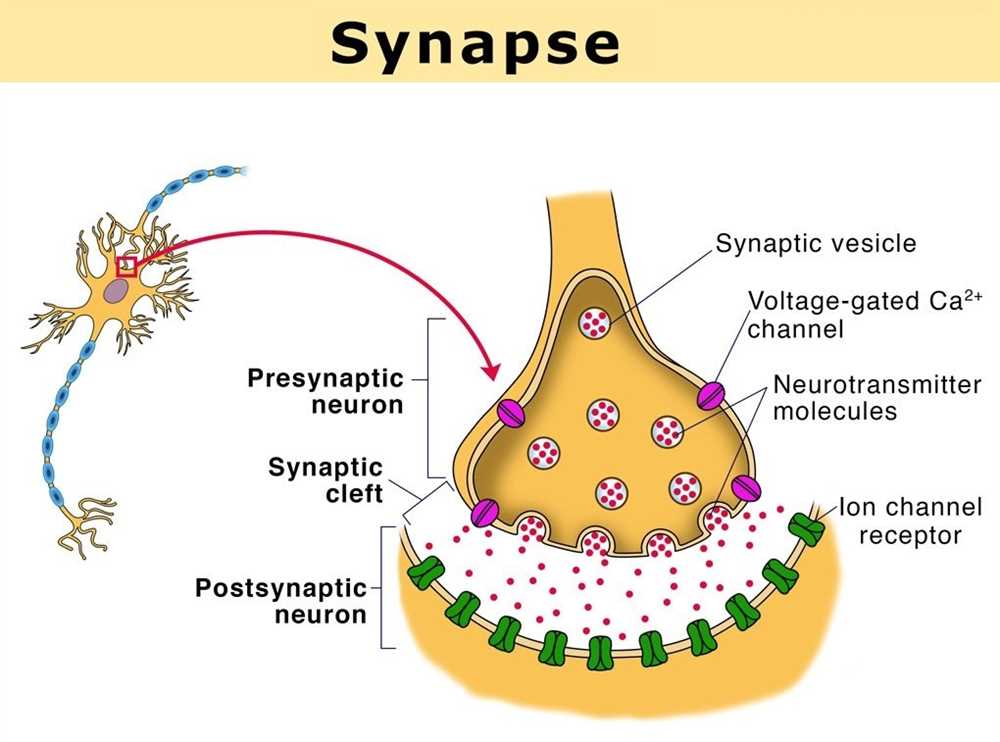
Pre-Synaptic Neuron: The pre-synaptic neuron is the sender of the signal. It contains the cell body, dendrites, and axon. The axon carries the electrical impulse known as an action potential from the cell body to the synapse.
Synaptic Cleft: The synaptic cleft is the small gap between the pre-synaptic neuron and the post-synaptic neuron. It is filled with extracellular fluid and acts as a barrier for the diffusion of neurotransmitters.
Post-Synaptic Neuron: The post-synaptic neuron is the receiver of the signal. It contains dendrites that receive the neurotransmitters released by the pre-synaptic neuron. These neurotransmitters bind to receptors on the post-synaptic neuron, leading to the generation of a new electrical impulse.
Neurotransmitters: Neurotransmitters are the chemical messengers that transmit signals across the synapse. Common neurotransmitters include dopamine, serotonin, and acetylcholine. They are stored in vesicles in the pre-synaptic neuron and are released into the synaptic cleft upon the arrival of an action potential.
Receptors: Receptors are proteins located on the post-synaptic neuron that bind with specific neurotransmitters. These receptors trigger changes in the post-synaptic neuron, allowing the transmission of the signal.
Neurotransmitter Reuptake: After the neurotransmitters have transmitted the signal, some of them are taken back up into the pre-synaptic neuron in a process called reuptake. This allows for recycling and reusing of the neurotransmitters.
Excitatory and Inhibitory Synapses: Synapses can either be excitatory or inhibitory, depending on the type of neurotransmitter released. Excitatory synapses increase the likelihood of the post-synaptic neuron firing, while inhibitory synapses decrease the likelihood of firing.
The Anatomy of Synapse Worksheet provides a comprehensive understanding of the different components and processes involved in synaptic transmission. It is an essential tool for neuroscience students and researchers to grasp the intricacies of synaptic communication in the brain.
Understanding the Synapse: Definition and Function
The synapse is a crucial component of the nervous system, responsible for transmitting information between neurons. It is the point where two neurons meet and communicate with each other. This communication occurs through the release and reception of chemical signals, known as neurotransmitters. The synapse plays a vital role in the transmission of electrical impulses and the integration of information within the brain.
Definition: A synapse is a junction between two nerve cells, where electrical signals are transmitted through the release of neurotransmitters.
Function: The primary function of the synapse is to enable communication between neurons. When an electrical impulse reaches the end of a neuron, it triggers the release of neurotransmitters into the synapse. These neurotransmitters then bind to receptors on the adjacent neuron, transmitting the signal across the synapse. This process allows information to be passed from one neuron to another, allowing for the coordination of complex behaviors and bodily functions.
The synapse can be classified into two main types: chemical synapses and electrical synapses. In chemical synapses, the transmission of information occurs through the release and binding of neurotransmitters. This type of synapse is the most common in the human brain. On the other hand, electrical synapses involve direct electrical connections between neurons, allowing for rapid communication.
In conclusion, the synapse is a vital component of the nervous system, enabling communication between neurons through the release and reception of chemical signals. Understanding the anatomy and function of the synapse is essential in furthering our knowledge of how the brain functions and how it processes information.
Structure of a Synapse: Key Components
A synapse is a specialized structure that allows the transmission of signals between neurons. It is composed of several key components that play different roles in the communication process.
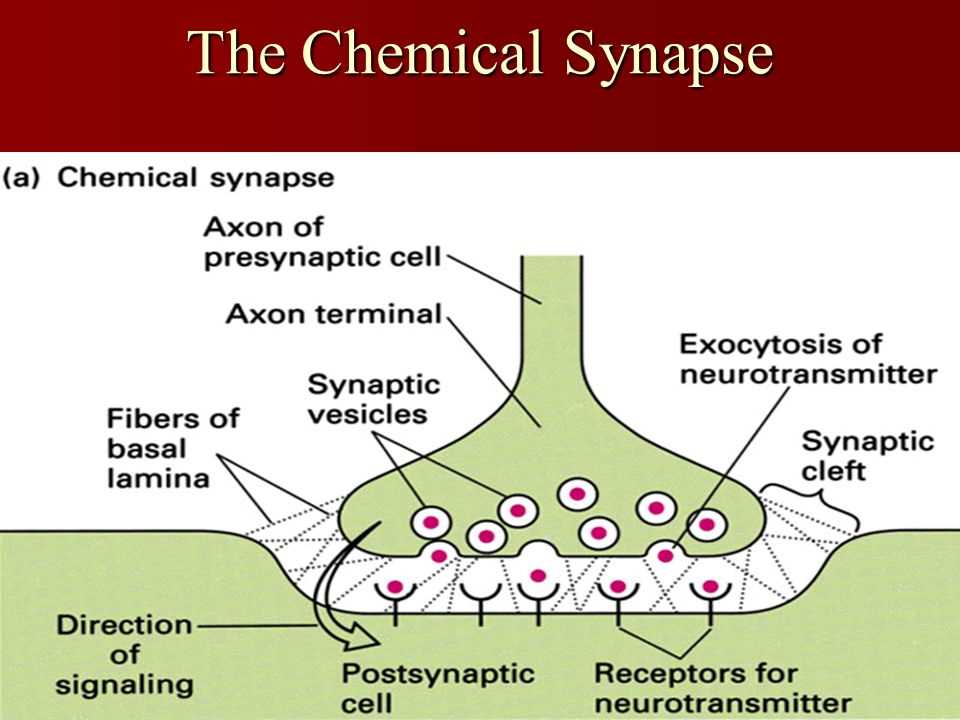
1. Presynaptic Terminal: The presynaptic terminal is the end of the neuron that sends the signal. It contains synaptic vesicles, which store neurotransmitters, and the active zones, where neurotransmitter release occurs. These two components work together to transmit the electrical signal into a chemical signal.
2. Synaptic Cleft: The synaptic cleft is the narrow gap between the presynaptic and postsynaptic membranes. It is filled with extracellular fluid and acts as a barrier between the two neurons. The neurotransmitters released from the presynaptic terminal diffuse across the synaptic cleft to reach the postsynaptic membrane.
3. Postsynaptic Membrane: The postsynaptic membrane is the end of the neuron that receives the signal. It contains receptor proteins that bind to neurotransmitters and trigger changes in the postsynaptic neuron. These changes can lead to the generation of an electrical signal called a post-synaptic potential.
4. Synaptic Vesicles: Synaptic vesicles are small sacs located in the presynaptic terminal. They store neurotransmitters, which are chemicals that transmit signals between neurons. When an electrical signal reaches the presynaptic terminal, the synaptic vesicles release the neurotransmitters into the synaptic cleft.
5. Neurotransmitters: Neurotransmitters are chemical messengers that transmit signals between neurons. They are released from the synaptic vesicles into the synaptic cleft and bind to receptor proteins on the postsynaptic membrane. Different neurotransmitters have different effects on the postsynaptic neuron, and their release and binding are tightly regulated to ensure proper communication between neurons.
6. Receptor Proteins: Receptor proteins are located on the postsynaptic membrane. They bind to specific neurotransmitters and initiate a response in the postsynaptic neuron. The binding of neurotransmitters to receptor proteins can activate or inhibit the postsynaptic neuron, depending on the type of receptor and neurotransmitter involved.
Overall, the structure of a synapse is complex and involves multiple components working together to transmit signals between neurons. Understanding the key components of a synapse is essential for studying the communication and functioning of the nervous system.
Neurons and Neurotransmitters: Their Role in Synaptic Communication
Neurons, also known as nerve cells, are the fundamental building blocks of the nervous system. They are specialized cells that transmit information throughout the body. The brain, spinal cord, and peripheral nerves are all composed of neurons.
Neurons communicate with each other through tiny gaps called synapses. These synapses allow for the transmission of electrical signals from one neuron to another. Neurotransmitters play a crucial role in this process, as they are the chemical messengers that carry the signals across the synapse.
Neurotransmitters are small molecules that are released by one neuron and bind to receptors on another neuron. They can either excite the receiving neuron, making it more likely to generate an action potential, or inhibit it, making it less likely to generate an action potential. This excitatory and inhibitory balance is crucial for proper synaptic communication.
Examples of neurotransmitters include:
- Acetylcholine: involved in muscle movement, learning, and memory.
- Dopamine: plays a role in motivation, reward, and pleasure.
- Serotonin: regulates mood, sleep, and appetite.
- GABA: an inhibitory neurotransmitter that helps to calm the brain.
| Neurotransmitter | Function |
|---|---|
| Acetylcholine | Involved in muscle movement, learning, and memory. |
| Dopamine | Plays a role in motivation, reward, and pleasure. |
| Serotonin | Regulates mood, sleep, and appetite. |
| GABA | An inhibitory neurotransmitter that helps to calm the brain. |
The release and uptake of neurotransmitters are tightly regulated processes that ensure proper synaptic communication. When there is an imbalance in neurotransmitter levels or functioning, it can lead to various neurological disorders, including depression, anxiety, and Parkinson’s disease.
In conclusion, neurons and neurotransmitters play a crucial role in synaptic communication. They allow for the transmission of signals between neurons, which is essential for the functioning of the nervous system. The balance of excitatory and inhibitory neurotransmitters is vital for proper brain function and mental health.
Synaptic Transmission: How Signals Are Sent and Received
In the complex network of the brain, communication between neurons happens through a process called synaptic transmission. Synapses are the junctions between neurons where chemical signals are transmitted from one neuron to another. This is a crucial process that allows information to be passed from one part of the brain to another.
When an electrical signal, or action potential, reaches the end of a neuron, it triggers the release of tiny chemical messengers called neurotransmitters into the synapse. These neurotransmitters diffuse across the synapse and bind to specialized receptors on the surface of the receiving neuron. The binding of the neurotransmitter to its receptor causes a series of biochemical events within the receiving neuron, ultimately leading to either the generation of a new action potential or the inhibition of further signaling.
This process of synaptic transmission is highly regulated and can be modified in order to fine-tune the communication between neurons. For example, the amount of neurotransmitter released into the synapse can be adjusted based on the needs of the neural circuit. Additionally, the strength of the connection between two neurons can be modified through a process called synaptic plasticity, which is thought to underlie learning and memory.
Overall, synaptic transmission is a fundamental process in the brain that allows for the transmission of signals and the integration of information. Understanding the intricacies of this process is crucial for unraveling the complexities of the brain and developing treatments for neurological disorders.
The Role of Ion Channels in Synaptic Signaling
In synaptic signaling, the communication between neurons relies on the transmission of electrical signals through synapses. Ion channels play a crucial role in this process by allowing the flow of ions across the neuronal membrane, which generates and modulates the electrical signals.
Ion channels: Ion channels are specialized proteins that are embedded in the neuronal membrane. They have a pore through which ions can pass, and their opening and closing are regulated by various factors, including voltage changes and neurotransmitter binding.
There are several types of ion channels involved in synaptic signaling. The two main types are voltage-gated ion channels and ligand-gated ion channels.
- Voltage-gated ion channels: These channels open or close in response to changes in the electrical potential across the neuronal membrane. When the membrane potential reaches a certain threshold, the voltage-gated ion channels open, allowing ions to flow in or out of the neuron. This generates an action potential, the electrical signal that travels along the neuron.
- Ligand-gated ion channels: These channels open or close in response to the binding of specific neurotransmitters. When a neurotransmitter binds to a ligand-gated ion channel, it causes the channel to open, allowing ions to flow through. This can lead to either depolarization (excitatory response) or hyperpolarization (inhibitory response) of the neuron, depending on the type of neurotransmitter and ion involved.
The opening and closing of ion channels is a key mechanism in synaptic signaling. It allows for the integration of multiple inputs from different neurons and the precise control of neuronal activity. Understanding the role of ion channels in synaptic signaling is essential for unraveling the complexities of brain function and for developing treatments for neurological disorders.
Synaptic Plasticity: The Ability of Neurons to Change
Synaptic plasticity is a fundamental property of the nervous system that allows neurons to adapt and change in response to an individual’s experiences and the demands of the environment. It is the ability of synapses, the connections between neurons, to strengthen or weaken in response to activity patterns and input. This dynamic nature of synapses underlies learning and memory formation, as well as other cognitive processes.
At the cellular level, synaptic plasticity involves changes in the strength of the synaptic connection, known as synaptic strength or synaptic efficacy. These changes are mediated by various molecular and cellular mechanisms, including alterations in neurotransmitter release, receptor expression, and dendritic spine morphology. The balance between these mechanisms determines whether a synapse becomes stronger or weaker, facilitating or inhibiting the transmission of signals between neurons.
One of the key forms of synaptic plasticity is called long-term potentiation (LTP), which refers to the long-lasting strengthening of synaptic connections. LTP is thought to be the cellular basis of learning and memory, as it allows for the modification and storage of information in the brain. Conversely, long-term depression (LTD) is the weakening of synapses and is also involved in shaping neural circuits and information processing.
Importantly, synaptic plasticity is not a static phenomenon, but rather a dynamic process that occurs throughout life. It plays a crucial role in brain development, allowing for the formation and refinement of neural circuits during critical periods. Additionally, synaptic plasticity continues to shape the adult brain, enabling learning, memory consolidation, and adaptation to changes in the environment.
In conclusion, synaptic plasticity is the ability of neurons to change their connections and strength in response to experience and activity. It is a fundamental mechanism underlying learning, memory, and neural development. By understanding the intricacies of synaptic plasticity, scientists hope to uncover new insights into brain function and develop interventions for neurological disorders.