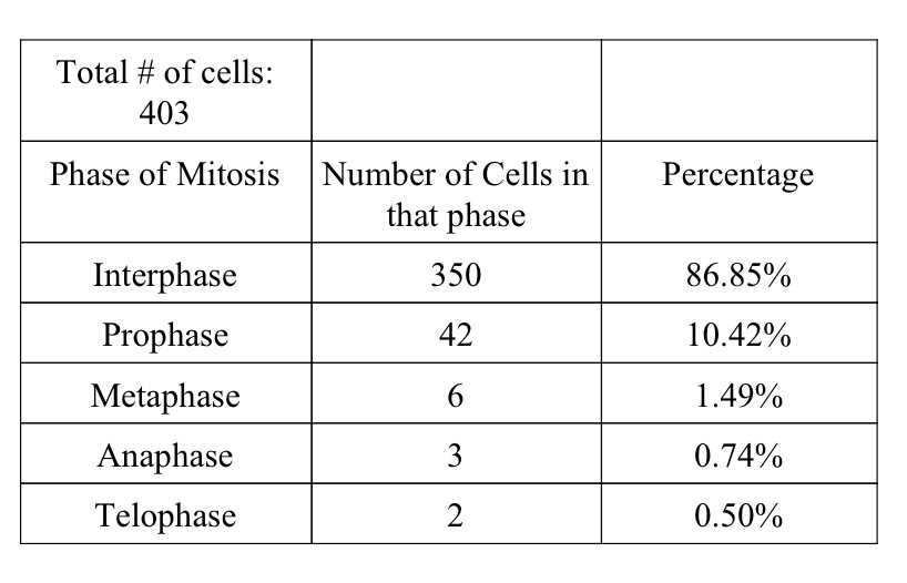
Answer key for the mitosis lab is crucial for understanding the process of cell division. This lab helps students grasp the concept of mitosis and its various stages. By observing and analyzing prepared microscope slides, students can identify the different phases of mitosis and understand the importance of this process in the growth and development of organisms.
The mitosis lab answer key provides students with a guide to identifying and labeling the different stages of mitosis, including interphase, prophase, metaphase, anaphase, and telophase. It also includes explanations and descriptions of each stage, helping students understand the changes that occur in the nucleus and cytoplasm during mitosis.
With the help of the answer key, students can compare their observations with the correct answers to ensure accuracy and deepen their understanding of mitosis. This lab also introduces students to the importance of proper scientific observation and recording data, as they are required to record their observations and label them correctly.
Overall, the mitosis lab answer key serves as an essential tool for students to learn about mitosis and practice their skills in observation and recording data. It allows students to connect theoretical knowledge with practical application, ultimately enhancing their understanding of cell division and its significance in biological processes.
What is Mitosis?
Mitosis is a fundamental process of cell division that occurs in eukaryotic cells. It is responsible for the growth, development, and maintenance of multicellular organisms. During mitosis, a single cell divides into two genetically identical daughter cells, each containing the same number of chromosomes as the parent cell. This process plays a crucial role in tissue repair, organ regeneration, and the production of new cells.
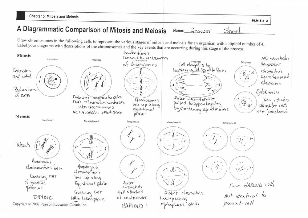
The process of mitosis is divided into four distinct phases: prophase, metaphase, anaphase, and telophase. During prophase, the chromosomes condense, the nuclear envelope breaks down, and the mitotic spindle forms. In metaphase, the chromosomes align along the equator of the cell. During anaphase, the sister chromatids separate and move towards opposite poles of the cell. Finally, in telophase, the chromosomes decondense, the nuclear envelope reforms, and the cell undergoes cytokinesis, resulting in the formation of two daughter cells.
Mitosis is a highly regulated process that ensures the accurate distribution of genetic material to each daughter cell. It is controlled by a complex network of proteins and signaling pathways that coordinate the events of each phase. Any disruption or errors in mitosis can lead to genetic instability, chromosomal abnormalities, and diseases such as cancer.
Overall, mitosis is a vital process that allows organisms to grow, repair damaged tissues, and maintain their cellular integrity. Through the precise division of genetic material, mitosis ensures the proper distribution of chromosomes and the continuity of life.
Why is studying mitosis important?
Studying mitosis is crucial for understanding the fundamental processes of cell division and growth. Mitosis is the process by which cells divide and reproduce, allowing organisms to grow and repair damaged tissues. It is a highly regulated and tightly controlled process that ensures the proper distribution of genetic material to the resulting daughter cells.
Firstly, studying mitosis helps researchers and scientists gain insights into the mechanisms and regulation of cell division. By understanding the intricate steps involved in mitosis, scientists can unravel the complex molecular events that govern cell proliferation. This knowledge is essential in the field of cancer research, as malfunctioning mitosis can lead to uncontrolled cell growth and tumor formation.
Secondly, studying mitosis is important in developmental biology. During embryonic development, cells undergo rapid and coordinated growth through mitosis. By studying mitosis in different organisms, researchers can gain a better understanding of how tissues and organs form and develop. This knowledge is essential for studying birth defects and finding potential treatments.
Furthermore, studying mitosis allows for better comprehension of genetic inheritance and evolution. Proper distribution of genetic material during mitosis ensures that each daughter cell receives a complete set of chromosomes. Any errors or abnormalities during this process can lead to genetic disorders or mutations. By studying mitosis, scientists can uncover the causes of genetic diseases and explore potential genetic therapies.
In conclusion, studying mitosis plays a crucial role in advancing our understanding of cell division, growth, development, and genetic inheritance. It provides valuable insights into the fundamental processes of life and has significant implications for fields such as cancer research, developmental biology, and genetics. Without a thorough understanding of mitosis, many aspects of biology and medicine would remain enigmatic.
Understanding the process of mitosis
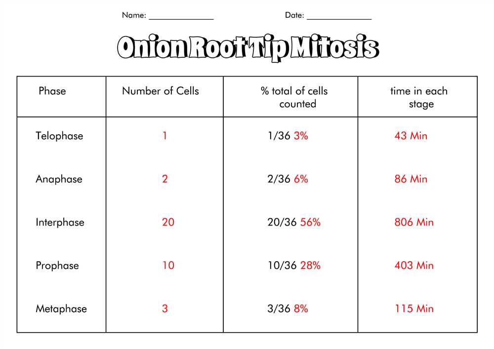
Mitosis is a fundamental process in cell division, allowing the growth and development of organisms and the replacement of old or damaged cells. It is a highly regulated and precise process that ensures the distribution of genetic material to daughter cells. By understanding the steps and mechanisms involved in mitosis, scientists can gain valuable insights into the maintenance and functioning of organisms.
The process of mitosis can be divided into several distinct phases: prophase, prometaphase, metaphase, anaphase, and telophase. Each phase is characterized by specific events and changes in cellular structure. During prophase, the genetic material condenses into chromosomes, and the nuclear envelope disintegrates. In prometaphase, the spindle fibers attach to the chromosomes, while the nuclear envelope continues to break down. Metaphase is marked by the alignment of chromosomes along the equator of the cell. In anaphase, the sister chromatids separate and move towards opposite poles of the cell. Finally, during telophase, the nuclear membrane reforms, and the chromosomes decondense.
The regulation and significance of mitosis
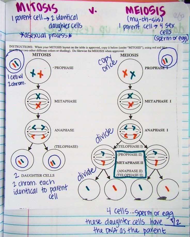
The process of mitosis is tightly regulated to ensure accurate and faithful distribution of genetic material. Various proteins and checkpoints monitor key events during mitosis, ensuring that each step is completed correctly before proceeding to the next. Mistakes or errors in mitosis can lead to chromosomal abnormalities and genetic disorders.
Mitosis is a crucial process for the growth, development, and repair of organisms. It is responsible for the formation of new cells during embryonic development and the regeneration of tissues in adult organisms. Additionally, mitosis plays a vital role in the maintenance of healthy tissues and the replacement of damaged or dying cells. By understanding the intricate mechanisms of mitosis, scientists can develop targeted therapies for diseases characterized by abnormal cell division, including cancer.
The Stages of Mitosis
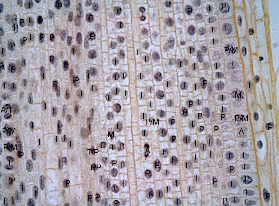
Mitosis is the process by which a single cell divides into two identical daughter cells. This cell division is necessary for growth, repair, and reproduction in multicellular organisms. Mitosis consists of several distinct stages, each with its own unique characteristics and functions.
The first stage of mitosis is called prophase. During prophase, the replicated DNA condenses into tightly coiled structures called chromosomes. The nuclear envelope also begins to break down, allowing the chromosomes to become more accessible. Additionally, spindle fibers form and attach to the centromeres of each chromosome.
The next stage is metaphase. In metaphase, the chromosomes align themselves along the equator of the cell, known as the metaphase plate. The spindle fibers help to align the chromosomes accurately, ensuring that each daughter cell receives the correct number of chromosomes. This alignment is crucial for the successful division of genetic material.
Anaphase follows metaphase. During anaphase, the sister chromatids of each chromosome separate and move towards opposite ends of the cell. This separation is facilitated by the shortening of the spindle fibers, which pull the chromatids towards the poles. As the chromatids move apart, the cell elongates and prepares for cytokinesis.
The final stage of mitosis is telophase. Telophase is characterized by the reformation of the nuclear envelope around the separated chromosomes. The chromosomes also begin to decondense, returning to their extended state. At this point, cytokinesis occurs, dividing the cytoplasm and organelles between the two daughter cells. This results in the formation of two identical daughter cells, each with its own complete set of chromosomes.
In conclusion, mitosis is a complex and highly regulated process that ensures the accurate division of genetic material. Each stage of mitosis plays a critical role in this process, from the condensation and alignment of chromosomes to the separation of sister chromatids and the final formation of two daughter cells. Understanding these stages is essential for studying cell division and the processes that regulate it.
Cell division and chromosome replication
Cell division is a vital process in the life cycle of living organisms. It allows organisms to grow, repair damaged tissues, and reproduce. One type of cell division is called mitosis, which involves the replication and distribution of chromosomes to daughter cells.
Chromosome replication is an essential step in mitosis. Before a cell can divide, it must first duplicate its genetic material to ensure that each daughter cell receives a complete set of chromosomes. This replication process occurs during the S phase of the cell cycle, where the DNA strands unwind and separate, allowing new complementary strands to be synthesized.
During chromosome replication, each DNA molecule acts as a template for the synthesis of a new complementary strand. This process is facilitated by enzymes called DNA polymerases, which add nucleotides to the growing DNA chain. As the replication proceeds, two identical copies of each chromosome are formed, known as sister chromatids.
Once the chromosomes have been successfully replicated, cell division can proceed. During mitosis, the sister chromatids are separated and distributed to opposite ends of the cell, ensuring that each daughter cell receives an equal number of chromosomes. This process is followed by cytokinesis, where the cell membrane pinches in and divides the cytoplasm, resulting in the formation of two genetically identical daughter cells.
In conclusion, cell division and chromosome replication are essential processes for the growth, development, and reproduction of organisms. These processes ensure the accurate distribution of genetic material and the formation of genetically identical daughter cells.
Conducting the mitosis lab
Conducting the mitosis lab is an essential part of understanding the process of cell division. This lab allows students to observe and analyze the different stages of mitosis in plant and animal cells.
The first step in conducting the mitosis lab is to prepare the slides for observation. Plant and animal cells are typically collected from tissues such as the root tip or the tip of an onion. These cells are then carefully placed on a microscope slide using a pipette or a dropper. A cover slip is added on top of the cells to protect them and ensure clarity during observation.
Once the slides are ready, students can begin the observation and analysis of the cells under a microscope. They can use a light microscope or a binocular microscope, depending on the availability and the preferences of the teacher. The cells are observed at different magnifications to clearly identify the different stages of mitosis, such as interphase, prophase, metaphase, anaphase, and telophase.
During the lab, students are encouraged to make detailed observations and sketch the cells at each stage of mitosis. They may also measure the durations of each phase and calculate the percentage of cells in each stage. This analysis helps them understand the dynamic nature of mitosis and how it contributes to cell growth and reproduction.
In conclusion, conducting the mitosis lab provides students with a hands-on experience of observing and analyzing cell division. It helps them gain a deeper understanding of the process and its significance in biological systems. This lab also hones their skills in microscopy, data analysis, and scientific observation, making it a valuable learning activity in the field of biology.
Equipment and Materials Needed
For the mitosis lab, it is important to have the right equipment and materials to properly observe and analyze the process. This will ensure accurate results and a comprehensive understanding of mitosis. Below is a list of essential items:
1. Microscope: A high-quality microscope is crucial for observing the cells and their division during mitosis. Make sure to have a microscope that offers a magnification of at least 400x to clearly view the cellular structures.
2. Slides: Glass slides are used to hold the specimens under the microscope. Prepare enough slides to place your samples for examination and analysis.
3. Cover slips: Cover slips are placed on top of the specimens on the slides to protect them and prevent contamination. Use cover slips that are the appropriate size for your slides.
4. Specimens: Obtain prepared slides that contain cells undergoing mitosis. These slides can be purchased from biological supply companies or prepared in advance using suitable cell cultures or tissue samples.
5. Staining agents: To enhance the visibility of cellular structures, staining agents such as methylene blue or iodine solution may be used. Follow the instructions provided with the staining agent and use it cautiously.
6. Timer or clock: To accurately record the duration of each stage of mitosis, a timer or clock is necessary. This will help ensure that you capture the data needed for analysis.
7. Lab notebook: Keep a detailed lab notebook to record observations, data, and any notes related to the procedure. This will help organize your findings and serve as a reference for future analysis.
Obtaining and organizing the necessary equipment and materials in advance will save time and ensure a smooth execution of the mitosis lab. Prepare your work area accordingly, ensuring that it is clean and well-lit to facilitate accurate observation and analysis.