
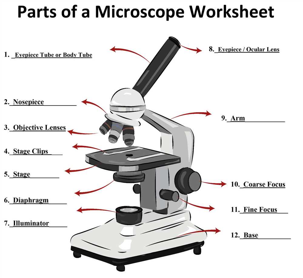
Microscopy has long been a vital tool in biology and medical research, enabling scientists to observe and analyze the intricate details of living organisms. However, traditional microscopes have their limitations, such as the need for expensive equipment and the inability to easily share and collaborate on observations. The emergence of virtual microscopy has revolutionized the field, providing a new way for researchers to explore and understand the complex world of biology.
With the advent of virtual microscopy, scientists are now able to access a digital platform that simulates the experience of using a conventional microscope. This innovative technology allows users to magnify, pan, and zoom on digital samples with the same level of precision and accuracy as a physical microscope. By utilizing high-resolution imaging and advanced algorithms, the virtual microscope offers unprecedented clarity and detail, unravelling the secrets of biological structures at the cellular and molecular level.
One of the most significant advantages of the virtual microscope is its ability to facilitate collaboration among researchers. Unlike traditional microscopes, which limit observations to a single person, the virtual microscope enables multiple scientists to simultaneously view and annotate samples remotely. This feature opens up new possibilities for interdisciplinary research and global collaboration, as scientists from different regions can easily exchange ideas and analyze data together, breaking down the barriers of physical location.
What is a Virtual Microscope?
A virtual microscope is a technological tool that allows users to examine and study microscopic samples digitally, without the need for physical slides or traditional microscopes. It uses advanced imaging techniques and software to create a virtual representation of the sample, which can be viewed, manipulated, and analyzed on a computer or other digital device. This technology has revolutionized the field of microscopy by providing researchers, scientists, and students with a convenient and efficient way to explore and study microscopic structures.
Virtual microscopes offer several advantages over traditional microscopes. Firstly, they eliminate the need for physical specimen preparation and storage, as digital samples can be easily accessed and shared online. This makes it easier for researchers in different locations to collaborate and exchange information. Additionally, virtual microscopes often provide enhanced imaging capabilities, such as the ability to zoom in and out, adjust the lighting and contrast, and even generate 3D images. These features allow for more detailed and accurate analysis of microscopic structures.
Key Features of a Virtual Microscope:
- Digitization: The process of converting physical microscope slides into digital images.
- Virtual Slides: Digital representations of microscope slides that can be viewed and manipulated on a computer.
- Image Analysis: Software tools that enable users to measure, annotate, and analyze the digital images.
- Remote Access: The ability to access and view virtual slides from anywhere, using an internet-connected device.
- Collaboration: Features that allow multiple users to view and discuss virtual slides simultaneously, promoting collaboration and knowledge sharing.
- Educational Resources: Virtual microscopes often come with educational materials, such as interactive tutorials, quizzes, and reference guides, to aid in learning and understanding of microscopic structures.
How does a Virtual Microscope Work?

A virtual microscope is a powerful tool that allows users to view and analyze microscopic specimens from the comfort of their computer screens. This technology has revolutionized the field of microscopy and has made it possible for researchers and students to access and study samples without the need for traditional microscope equipment.
So how does a virtual microscope work? Instead of using physical lenses to magnify the image, a virtual microscope relies on digital imaging technology. The specimen is first prepared and mounted on a glass slide, just like with a traditional microscope. However, instead of placing the slide on a microscope stage, it is scanned and digitized using a high-resolution camera or scanner.
The digital image is then uploaded to a computer software program, which allows the user to manipulate and navigate the image in various ways. Using the software, the user can zoom in and out, adjust the focus, and even change the contrast and brightness of the image. This level of control and customization is one of the key advantages of virtual microscopes.
In addition to the image manipulation features, virtual microscopes often have annotation tools that allow users to mark and label specific parts of the specimen. This can be particularly useful for educational purposes, as it allows teachers and students to easily identify and discuss specific structures or features.
Virtual microscopes also offer the option to save and share images, making it easy to collaborate and communicate findings with others. In some cases, virtual microscopes can even be connected to a network, allowing multiple users to access and view the same specimen simultaneously.
Overall, a virtual microscope provides a convenient and flexible alternative to traditional microscopy. It allows for remote access to specimens, offers powerful image manipulation tools, and promotes collaboration and sharing within the scientific community.
Advantages of a Virtual Microscope over Traditional Microscopes
In recent years, virtual microscopes have emerged as a powerful tool in the field of microscopy, offering several advantages over traditional microscopes. One of the main advantages is the ability to access and analyze digital images of specimens remotely, without the need for physical slides. This allows researchers and students to study samples from anywhere in the world, saving time and costs associated with shipping and handling of delicate samples.
Additionally, virtual microscopes provide enhanced convenience and flexibility. With a traditional microscope, users are limited by the physical limitations of the instrument, such as the size of the stage and the number of available objective lenses. In contrast, virtual microscopes offer the ability to zoom in and out, change magnification levels, and manipulate images to highlight specific details. This flexibility allows for more precise analysis and makes it easier to share findings with colleagues or students.
Another advantage of virtual microscopes is the ability to capture and store high-resolution images of specimens. Traditional microscopes often rely on the user’s ability to accurately observe and sketch what they see. However, human error and limitations can result in inaccuracies or inconsistencies in documentation. With virtual microscopes, images can be captured digitally, ensuring that all details are accurately recorded and can be easily accessed at a later time if needed.
Furthermore, virtual microscopes offer advanced features, such as measurement tools, annotations, and the ability to overlay multiple images. These features allow for more comprehensive analysis and facilitate the comparison of different samples or time points. In addition, virtual microscopes often incorporate automated image analysis algorithms, which can help researchers identify and quantify features of interest more efficiently.
In conclusion, virtual microscopes offer numerous advantages over traditional microscopes. They provide remote access, enhanced convenience and flexibility, accurate documentation, and advanced analytical features. As technology continues to advance, virtual microscopes are likely to become increasingly vital in the field of microscopy and contribute to significant advancements in research and education.
Applications of Virtual Microscopes
Virtual microscopes have revolutionized the field of microscopy by providing researchers with a powerful tool to study specimens in a virtual environment. These virtual microscopes have a wide range of applications in various scientific fields.
Education and training: Virtual microscopes have been widely used in educational institutions to enhance the learning experience of students. They provide a realistic simulation of a traditional microscope, allowing students to practice their microscopy skills and explore different types of specimens without the need for physical samples. Virtual microscopes also enable educators to create interactive and engaging teaching materials, making it easier to explain complex scientific concepts.
Research and analysis: Virtual microscopes are instrumental in the field of research and analysis. They allow researchers to examine specimens in high resolution and manipulate the images to extract valuable information. Virtual microscopes also offer various imaging techniques, such as fluorescence and phase contrast microscopy, which can be used to study specific features of specimens. Furthermore, virtual microscopes enable researchers to collaborate remotely and share their findings with colleagues around the world.
Medical diagnostics: Virtual microscopes have found applications in the field of medical diagnostics. They enable pathologists to scan and digitize tissue samples, creating virtual slides that can be viewed and analyzed remotely. This technology has revolutionized the field of pathology, allowing for faster and more efficient diagnosis of diseases. Virtual microscopes also offer advanced image analysis algorithms that can aid in the detection and classification of abnormal cells or tissues, providing valuable insights for medical professionals.
Quality control: Virtual microscopes are increasingly being used in industries where quality control is essential, such as pharmaceuticals and manufacturing. They allow for detailed inspection and analysis of products, ensuring that they meet the required standards. Virtual microscopes enable the detection of defects or irregularities in samples, leading to improved product quality and reduced waste. They also provide a comprehensive documentation system, allowing for easy tracking and analysis of data.
In conclusion, virtual microscopes have diverse applications in education, research, medical diagnostics, and quality control. They provide a versatile and powerful tool for studying specimens in a virtual environment, offering numerous benefits in terms of flexibility, accessibility, and data analysis.
Virtual Microscopes in Medical Science
In the field of medical science, virtual microscopes have revolutionized the way researchers and clinicians study and analyze microscopic samples.
Virtual microscopes are powerful tools that allow users to view high-resolution images of microscopic samples, such as cells, tissues, and microorganisms, through a computer screen. These digital microscopes provide an immersive and interactive experience, offering magnification and navigational capabilities that go beyond what traditional optical microscopes can offer.
One of the key advantages of using virtual microscopes in medical science is the ability to easily share and collaborate on images. With a virtual microscope, researchers can capture digital images of samples and share them with colleagues across the globe. This not only speeds up the research process, but also allows for real-time discussions and collaborations, leading to faster breakthroughs in medical science.
Furthermore, virtual microscopes offer advanced imaging techniques that enhance the ability to study and diagnose diseases. For example, digital pathology utilizes virtual microscopy to analyze tissue samples and make accurate diagnoses. Pathologists can zoom in and pan across the samples, examine specific areas of interest, and annotate the images for clearer communication. This technology has significantly improved diagnostic accuracy and has the potential to revolutionize the field of pathology.
In summary, virtual microscopes have greatly advanced medical science by providing researchers and clinicians with a more efficient and effective way to study microscopic samples. With their ability to easily share images, collaborate in real-time, and utilize advanced imaging techniques, virtual microscopes are shaping the future of medical research and diagnosis.
Virtual Microscopes in Research
Virtual microscopes have revolutionized the field of research, providing scientists with a powerful tool to study and analyze microscopic specimens. These advanced digital imaging systems allow researchers to view samples in high resolution and capture detailed images. Virtual microscopes have a wide range of applications in various fields, including biology, medicine, and materials science.
One of the main advantages of virtual microscopes is their ability to provide remote access. Scientists can now access and control microscopes from anywhere in the world, without the need for physical presence in the laboratory. This has greatly facilitated collaboration between researchers and enabled real-time data sharing and analysis. Additionally, virtual microscopes have eliminated the need for slides and physical samples, reducing the risk of contamination and saving valuable time and resources.
Virtual microscopes also offer advanced imaging capabilities, such as digital zoom, image stitching, and 3D visualization. These features allow researchers to explore specimens in detail, from multiple angles and at various magnifications. Virtual microscopes can also generate high-quality images that can be easily shared and analyzed using computer software. This has accelerated the research process and enabled more efficient data analysis.
In conclusion, virtual microscopes have transformed the way research is conducted by providing scientists with a powerful tool to study microscopic samples. The remote access, advanced imaging capabilities, and efficient data analysis offered by virtual microscopes have greatly enhanced collaboration and accelerated the research process. As technology continues to advance, virtual microscopes are likely to play an even more significant role in scientific research in the future.
Future Developments in Virtual Microscopy
Virtual microscopy is a rapidly advancing field with exciting potential for future developments. As technology continues to improve, the capabilities of virtual microscopes will only expand. One key area of future development is in the enhancement of image resolution and clarity. Higher resolution images will allow for more detailed examination and analysis of microscopic specimens, leading to greater accuracy in diagnosing diseases and identifying abnormalities.
Another area of future development is in the integration of artificial intelligence (AI) algorithms into virtual microscopy systems. AI algorithms can assist pathologists in analyzing and interpreting digital slides, helping to detect patterns and anomalies that may not be immediately apparent to the human eye. This integration of AI could revolutionize the field of pathology by streamlining the diagnostic process and improving accuracy.
In addition, virtual microscopy is likely to see advancements in its capabilities for data management and sharing. As more institutions and laboratories adopt virtual microscopy systems, there will be a need for standardized protocols for storing and transmitting digital slides. This will enable pathologists from different locations to easily access and share slides for consultations and collaborative research.
Furthermore, the development of virtual reality (VR) technology has the potential to transform the way we interact with microscopic specimens. Virtual reality can provide an immersive and interactive experience, allowing pathologists to explore and manipulate virtual slides in a three-dimensional environment. This could greatly enhance the learning and training opportunities for medical students and professionals, helping them develop a better understanding of microscopic structures and pathologies.
In summary, the future of virtual microscopy holds great promise. With advancements in image resolution, integration of AI algorithms, improved data management, and the incorporation of virtual reality technology, virtual microscopes will continue to revolutionize the field of pathology and enhance our understanding of the microscopic world.