
Gel electrophoresis is a common laboratory technique used to separate and analyze DNA, RNA, and proteins based on their size and charge. This technique has revolutionized the field of molecular biology and has become an essential tool in many research and clinical laboratories.
The basic principle of gel electrophoresis is that DNA, RNA, or proteins are loaded into wells at one end of a gel matrix. An electric current is then applied, causing the molecules to migrate through the gel towards the positive electrode. The migration rate of each molecule is determined by its size and charge, allowing for separation and analysis.
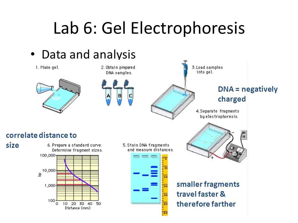
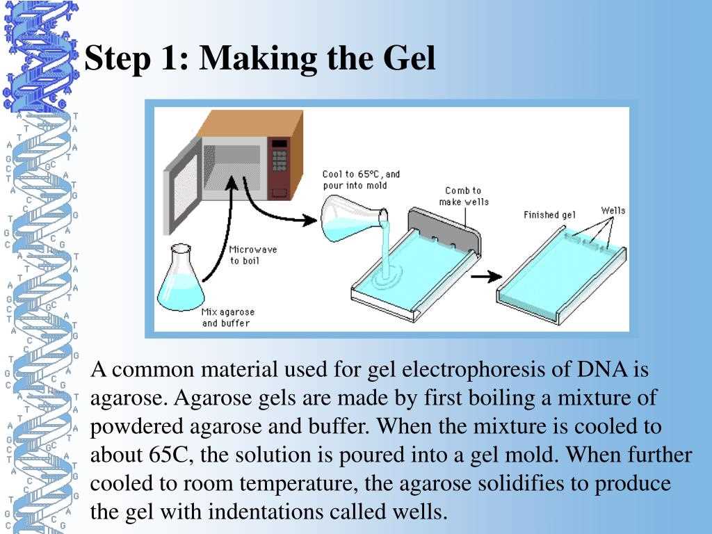
In a gel electrophoresis lab, students are typically provided with a DNA sample and are asked to analyze it using this technique. They first prepare an agarose gel, which is a polysaccharide derived from seaweed, and pour it into a gel tray. The gel is then submerged in a buffer solution that provides ions to conduct the electric current.
The students then load the DNA sample into wells on one end of the gel and connect the gel tray to a power supply. They run the gel at a specific voltage for a set amount of time, allowing the DNA fragments to migrate through the gel. After the run is complete, the students typically stain the gel with a DNA-binding dye, which allows them to visualize and analyze the separated DNA bands under UV light.
In this article, we will provide answers to common questions encountered in gel electrophoresis labs, including how to determine the size of DNA fragments, how to calculate the migration rate, and how to interpret the results. We will also discuss troubleshooting tips and variations of gel electrophoresis that are commonly used in different applications.
Gel Electrophoresis Lab Answers
In a gel electrophoresis lab, various DNA samples are analyzed using gel electrophoresis technique to determine their size and composition. Gel electrophoresis is a commonly used method in molecular biology and genetics research to separate and analyze DNA fragments based on their molecular weight.
The gel electrophoresis lab answers many questions related to DNA analysis. Firstly, it helps determine the size of DNA fragments by comparing their migration distance on a gel to DNA size markers of known lengths. By analyzing the pattern of DNA bands on the gel, scientists can estimate the size of unknown DNA fragments.
The gel electrophoresis lab also helps determine the composition of DNA samples. By using specific DNA stains or probes, scientists can visualize DNA bands on the gel and determine if the sample contains single-stranded or double-stranded DNA, RNA, or other DNA molecules. This information is crucial in understanding the nature and characteristics of the DNA sample being analyzed.
Additionally, the gel electrophoresis lab can provide insights into DNA mutations and genetic variations. By comparing DNA samples from different individuals or organisms, scientists can observe differences in band patterns, indicating variations in DNA sequences. This information can be used in genetic research or forensic analysis to identify individuals or study genetic diseases.
In conclusion, the gel electrophoresis lab is an essential tool in molecular biology and genetics research. It provides answers regarding the size, composition, and genetic variations of DNA samples. This information is valuable for understanding DNA structure, function, and its role in various biological processes.
What is Gel Electrophoresis?
Gel electrophoresis is a laboratory technique used to separate and analyze DNA, RNA, and proteins based on their size and charge. It is commonly used in molecular biology and biochemistry research as well as in forensic and clinical laboratories. The technique involves subjecting a mixture of molecules to an electric current, which causes them to move through a gel matrix towards a positively charged electrode.
The gel matrix used in gel electrophoresis is typically made of agarose or polyacrylamide. Agarose gels are most commonly used for separating DNA fragments, while polyacrylamide gels are preferred for the separation of smaller molecules such as RNA and proteins. The gel acts as a sieve, with smaller molecules moving more easily through the gel pores and thus traveling faster than larger molecules.
How does gel electrophoresis work?
Gel electrophoresis works based on the principle that DNA, RNA, and proteins have a net negative charge due to the phosphate groups or certain amino acids they contain. When an electric current is applied, the negatively charged molecules migrate through the gel towards the positive electrode. The movement of the molecules is influenced by their size and charge, resulting in the separation of different fragments or molecules according to their size.
To visualize the separated molecules, the gel is stained with a dye such as ethidium bromide or Coomassie blue, which binds to the DNA, RNA, or protein molecules and makes them visible under ultraviolet (UV) light or by staining with a colored reagent. The separated molecules can then be analyzed and studied further to understand their structure, function, or genetic information.
Applications of gel electrophoresis
Gel electrophoresis has a wide range of applications in various fields of biology and biochemistry. It is used for DNA fingerprinting, which is important in forensic analysis and paternity testing. The technique is also used in genetic research to study gene expression, genetic mutations, and DNA sequencing. In protein research, gel electrophoresis helps in analyzing protein size, purity, and identifying protein-protein interactions. Additionally, gel electrophoresis is used for the diagnosis of genetic disorders and infectious diseases in clinical laboratories.
- Advantages of gel electrophoresis:
- Relatively simple and cost-effective technique
- Allows for the separation and analysis of different molecules
- Can be used with various sample types, including DNA, RNA, and proteins
Principles and Techniques of Gel Electrophoresis
Gel electrophoresis is a commonly used technique in molecular biology to separate and analyze DNA, RNA, or proteins based on their size and charge. It is a fundamental tool in many laboratories and is essential for various applications, including DNA sequencing, DNA fingerprinting, and protein analysis. Gel electrophoresis works on the principle that charged molecules will migrate through a porous gel matrix when an electric field is applied.The process of gel electrophoresis involves several steps. First, a gel matrix is prepared by mixing a specific concentration of agarose or polyacrylamide with a buffer solution. Agarose gels are commonly used for DNA analysis, while polyacrylamide gels are used for protein analysis. The gel is cast in a horizontal or vertical apparatus and allowed to solidify.
Next, the samples to be analyzed are prepared. DNA or RNA samples are typically mixed with a loading dye that contains a tracking dye, such as bromophenol blue, and a dense solution, such as glycerol, which helps the sample sink into the wells. Protein samples are usually denatured and mixed with a loading buffer containing a reducing agent and a detergent to ensure even migration and proper separation.
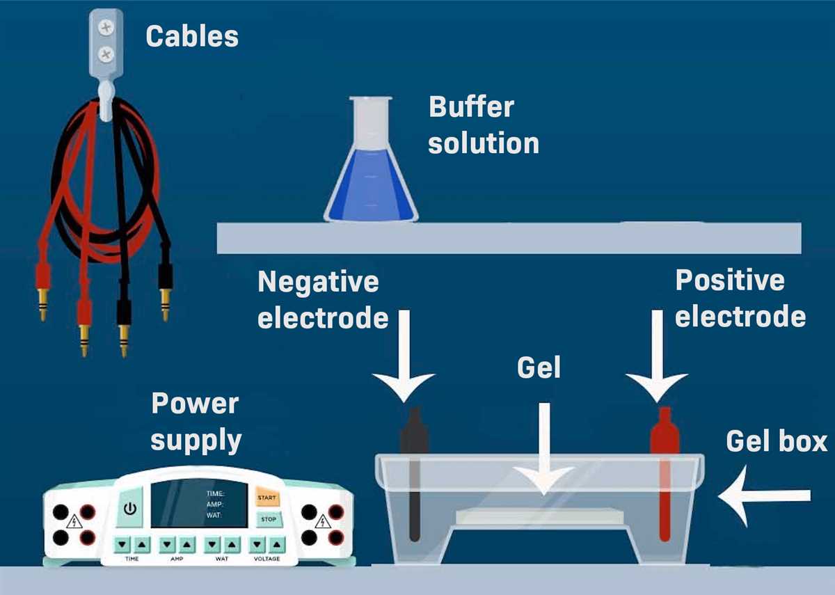
Once the gel and samples are ready, the gel is placed in an electrophoresis chamber filled with a buffer solution that provides ions for conductivity. The gel is carefully submerged in the buffer, and electrodes connected to a power supply are positioned at each end of the chamber. When the power supply is turned on, an electric current is applied, causing the negatively charged molecules to move towards the positive electrode, and the positively charged molecules to move towards the negative electrode.
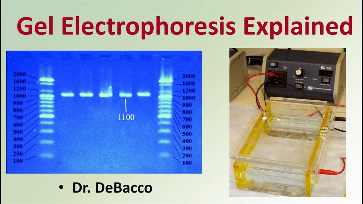
As the molecules move through the gel, they encounter resistance from the gel matrix, causing larger molecules to migrate more slowly than smaller ones. The gel acts like a sieving matrix, separating the molecules based on their size. After the electrophoresis run is complete, the gel is stained with a DNA-specific dye or a protein stain to visualize the separated molecules. The bands on the gel can be analyzed and compared to standard markers to determine the size or molecular weight of the molecules of interest.
In conclusion, gel electrophoresis is a powerful technique that allows for the separation and analysis of nucleic acids and proteins based on their size and charge. It is widely used in molecular biology laboratories and has revolutionized various fields, such as genetics, forensic science, and biomedical research. By understanding the principles and techniques of gel electrophoresis, scientists can gain valuable insights into the structure and function of biological molecules.
Materials and Methods for Gel Electrophoresis Lab
For the gel electrophoresis lab, several key materials and methods were utilized to separate and analyze DNA samples. The following provides an overview of the materials and methods used in the experiment.
Materials:
- Gel electrophoresis apparatus
- Agarose gel
- DNA samples
- DNA ladder markers
- Electrophoresis buffer
- Power supply
- Gel staining agent (e.g., ethidium bromide)
- Ultraviolet (UV) light source
In this lab, the gel electrophoresis apparatus was set up according to manufacturer instructions, with the agarose gel prepared by dissolving agarose powder in electrophoresis buffer. The resulting gel solution was poured into a gel tray and allowed to solidify. Meanwhile, DNA samples were prepared by adding a dye to the DNA to visualize it during electrophoresis.
After the gel had solidified, DNA samples and DNA ladder markers were loaded into separate wells in the gel. The gel tray was then placed into the gel electrophoresis apparatus, and the power supply was connected. A constant voltage was applied across the gel, causing the DNA samples to migrate through the gel towards the positive electrode. The gel was run for a predetermined amount of time to allow for separation of the DNA fragments.
Methods:
- Prepare the agarose gel by dissolving agarose powder in electrophoresis buffer.
- Pour the gel solution into a gel tray and allow it to solidify.
- Prepare DNA samples by adding a dye to visualize the DNA.
- Load the DNA samples and DNA ladder markers into separate wells in the gel.
- Place the gel tray into the gel electrophoresis apparatus.
- Connect the power supply and apply a constant voltage across the gel.
- Run the gel for a predetermined amount of time to allow for separation of the DNA fragments.
- Remove the gel tray from the apparatus and stain the gel with a staining agent.
- Visualize the DNA bands under a UV light source.
These materials and methods provided a framework for the gel electrophoresis lab, enabling the separation and analysis of DNA samples based on their size and charge. The experiment allowed for the detection of DNA fragments and provided valuable information for various scientific applications, such as genetic research and forensic analysis.
Results of Gel Electrophoresis Lab
During the gel electrophoresis lab, we conducted an experiment to separate and compare DNA samples based on their size and charge. The DNA samples used in the lab were obtained from different sources, such as bacteria, plants, and animals. By subjecting these samples to electrophoresis, we were able to observe the migration pattern and distinguish between the different DNA fragments.
The results of the gel electrophoresis lab showed distinct bands on the gel, indicating the separation of DNA fragments based on their size. Smaller DNA fragments migrated faster and traveled further in the gel, while larger fragments remained closer to the starting point. This allowed us to determine the relative sizes of the DNA fragments and compare them among the different samples.
By comparing the banding patterns on the gel, we were also able to analyze the similarities and differences between the DNA samples. For example, we could determine if two samples had similar DNA fragments or if one sample had additional or missing fragments compared to the others. This information can be used to explore genetic relationships, identify genetic mutations, or even determine the presence of specific genes.
In conclusion, the results of the gel electrophoresis lab provided valuable insights into the DNA samples we tested. The separation and analysis of DNA fragments based on their size and charge allowed us to compare different samples and observe their genetic characteristics. This technique is widely used in molecular biology research and has various applications in fields such as forensics, biotechnology, and medicine.
Interpretation of Gel Electrophoresis Lab Results
When analyzing the results of a gel electrophoresis lab, it is important to understand the purpose of the experiment and the expected outcomes. Gel electrophoresis is a commonly used technique in molecular biology that is used to separate and analyze DNA, RNA, or proteins based on their size and charge. The results obtained from the gel electrophoresis experiment can provide valuable information about the samples being analyzed.
One of the key aspects of interpreting gel electrophoresis lab results is understanding the concept of migration. During the electrophoresis process, DNA, RNA, or proteins are loaded into wells on the gel, and an electric current is applied. The molecules will migrate through the gel at different rates based on their size and charge. Smaller molecules will migrate faster than larger ones, resulting in distinct bands on the gel.
Interpreting the gel electrophoresis lab results involves analyzing the bands that appear on the gel. The position of each band corresponds to the migration of a specific molecule. By comparing the position of the bands to size markers or standards, one can estimate the size of the molecules being analyzed. The intensity of the bands also provides information about the concentration of the molecules.
Additionally, the presence or absence of specific bands can indicate the presence or absence of certain molecules or DNA fragments. For example, in a DNA gel electrophoresis lab, if a specific band appears in a sample but not in the control, it suggests that the sample contains a DNA fragment that is not present in the control. This can be particularly useful in genetic research and diagnostic applications.
In conclusion, interpreting the results of a gel electrophoresis lab involves analyzing the position, size, and intensity of the bands on the gel. This information can provide insights into the size, concentration, and presence/absence of specific molecules or DNA fragments. Understanding these results is crucial for drawing meaningful conclusions and advancing scientific research.
Applications and Importance of Gel Electrophoresis
Gel electrophoresis is a widely used technique in molecular biology that has numerous applications across various scientific fields. This technique allows scientists to separate and analyze DNA, RNA, and proteins based on their size and charge. Gel electrophoresis has revolutionized the field of genetics and has become an indispensable tool in many research laboratories.
Applications:
- Genetic research: Gel electrophoresis is extensively used in genetic research to study genetic disorders, genetic variation, and the organization of the genome. It enables scientists to analyze DNA fragments, detect mutations, and identify genetic markers.
- Forensic analysis: Gel electrophoresis plays a crucial role in forensic analysis by allowing scientists to compare DNA samples and identify suspects in criminal investigations. It is commonly used for DNA profiling and paternity testing.
- Medical diagnostics: Gel electrophoresis is instrumental in medical diagnostics for detecting and diagnosing diseases. It enables the identification of specific proteins or DNA fragments that are indicative of certain diseases, such as cancer or genetic disorders.
- Pharmaceutical development: Gel electrophoresis is utilized in the development and quality control of pharmaceutical drugs. It helps in analyzing and characterizing protein-based drugs, determining their purity, and detecting any impurities.
- Environmental monitoring: Gel electrophoresis is used in environmental monitoring to assess the presence of organisms or pollutants in environmental samples. For example, it can help identify the presence of specific bacteria in water samples or detect genetically modified organisms in food products.
Importance:
Gel electrophoresis is a powerful and versatile technique that has significantly advanced the fields of genetics, molecular biology, and biotechnology. Its importance lies in its ability to separate and analyze DNA, RNA, and proteins, providing crucial information about their structure, size, and abundance. Without gel electrophoresis, many important discoveries and advancements in the understanding of genetic disorders, forensic analysis, medical diagnostics, and drug development would not have been possible. This technique has revolutionized various scientific disciplines and continues to be an essential tool for scientists around the world.
Q&A:
What is gel electrophoresis?
Gel electrophoresis is a technique used to separate and analyze DNA, RNA, or proteins based on their size, charge, and shape.
What is the purpose of gel electrophoresis?
The purpose of gel electrophoresis is to separate DNA, RNA, or proteins into distinct bands or spots in order to analyze their composition, quantity, or purity.
How does gel electrophoresis work?
Gel electrophoresis works by placing a sample of DNA, RNA, or proteins into a gel matrix and applying an electric field. The samples migrate through the gel based on their charge, size, and shape, resulting in separation and the formation of distinct bands or spots.
What are the applications of gel electrophoresis?
Gel electrophoresis is used in various applications, such as DNA fingerprinting, genetic sequencing, gene expression analysis, paternity testing, and protein purification.
Why is gel electrophoresis important?
Gel electrophoresis is important because it allows scientists to analyze and study DNA, RNA, and proteins, which are essential molecules in biological research. It helps in understanding their structure, function, and interactions, and contributes to advancements in fields like medicine, genetics, and biotechnology.